A Case of Giant Becker’s Nevus
Keywords
LETTER
Sir,
Becker’s Nevus (BN) is a cutaneous hamartoma which is characterized by an unilateral, hyperpigmented patch with varying degrees of hypertrichosis [1]. We present a case of giant BN which is an extremely rare type of BN.
A 16-year-old Turkish female patient was admitted to our clinic with the complaint of a brown lesion located unilaterally on the right side of the body. The lesion had appeared when she was between 3-4 years old. Dermatological examination showed a wide brownish patch with well defined and irregular borders that spreaded from right ankle to ipsilateral middle trunk (Figure 1). Total involvement surface area was estimated to be around 3700cm2. There was hypertrichosis on the lesion on the lumbar area, and acneiform papules on the thigh (Figure 2). Histopathologic examination of the lesion revealed moderate acanthosis, hyperpigmentation of the basal layer and hyperplasia of the arrector pili muscle (Figure 3). Based on the history, physical examination and histopathological findings of the lesion, the patient was diagnosed as BN. The results of routine laboratory tests such as complete blood count, liver function tests and renal function tests were within normal limits. No pathologic finding was detected in X-ray examination of the legs and abdominal ultrasonography.
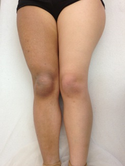
Figure 1: Wide hyperpigmented patch on the right lower extremity.
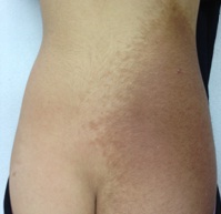
Figure 2: Hypertrichosis over the hyperpigmented patch on the right lumbar area.

Figure 3: Moderate acanthosis, hyperpigmentation of the basal layer, moderate flattening on rete ridges (H&E 200).
Prevalence of BN is nearly 0.52% [1]. BN usually has juvenile onset but, rarely, the cases with early onset or congenital cases have been reported [2]. In our case the lesion had started when the patient was 3 years old.
The abnormal androgen receptor activity may play a role in the pathogenesis of BN. Increased androgen receptor density has been shown via immunohistochemical staining, reverse transcriptase polymerase chain reaction of androgen receptor messenger RNA and ligand-binding assays in BN. Hypertrichosis and acneiform lesions on the BN may support the role of abnormal androgen receptor activity [3].
The most frequent localizations of the disease are trunk and legs. Also BN can be associated with BN syndrome. BN syndrome is characterized by BN with ipsilateral breast hypoplasia or other skin, skeletal and/or muscular disorders [4]. In our case there were no other pathological findings except BN.
An average size of BN is 125 cm2 [5]. There are few reports of giant BN in the literature. Any threshold size for giant BN has not been defined previously in literature. Reported cases of giant BN were patients who had usually bilateral torso involvement or a half of trunk [5-7]. BN has covered whole right lower extremity and ipsilateral middle trunk in our case. Khatami et al., reported a giant bilateral Becker's nevus in a 14 year old boy. The age of onset of the disease was when he was 8 years old [4]. Baeta et al., reported a case of giant BN syndrome with breast hypoplasia which had started at the age of 7 [7]. Our case is the 3rd case of giant BN and the age of onset of the lesion is earlier than the other reported cases.
Histologic evidences of BN are acanthosis, hyperkeratosis and hyperpigmentation of the basal layer. The number of melanocytes is generally normal or slightly increased and there are no nevus cells. In some BN cases, hamartomatous smooth muscle fibers may be seen on histopathology but we did not detect it on the histopatological examination of the lesion [7].
BN is only treated for cosmetic reasons. There are limited data about the use of laser for hypertrichosis and hyperpigmentation. Intense pulsed light laser and Erbium:YAG laser may be used for treatment [8]. Laser treatment was recommended to our patient.
We present this case because giant BN is a very rare type of Becker nevus and we want to emphasize that the diagnosis of BN should be kept in mind in patients with giant pigmented patches.
REFERENCES
- Ortonne JP, Bahadorian P, Fizpatrick TB et al. (2003) Hypomelanoses and hypermelanosis. In: Freedberg IM, Eisen AZ, Wolff K, Austen KF, Goldsmith LA, et al. (eds.). Fizpatrick's Dermatology in General Medicine. (6th edn), New York, Mc Graw-Hill. Pg: 836-880.
- Steiner D, Silva FA, Pessanha AC, Bialeski N, Feola C, et al. (2011) Do you know this syndrome? Becker nevus syndrome. An Bras Dermatol 86: 165-166.
- Kim YJ, Han JH, Kang HY, Lee ES, Kim YC (2008) Androgen receptor overexpression in Becker nevus: histopathologic and immunohistochemical analysis. J Cutan Pathol 35: 1121-1126.
- Khatami A, Seradj MH, Gorouhi F, Firooz A, Dowlati Y (2008) Giant bilateral becker nevus: a rare presentation. Pediatr Dermatol 25: 47-51.
- Issa G, Blalock TW, Lesher JL (2011) Patient with giant Becker's nevus and epidermal nevus. Dermatol Reports 3: 23.
- Crone AM, James MP (1997) Giant Becker's naevus with ipsilateral areolar hypoplasia and limb asymmetry. Clin Exp Dermatol 22: 240-241.
- Baeta IG, Viotti CV, Pereira AC, Costa Júnior SR, Bittencourt FV (2010) Becker's nevus syndrome: case report. An Bras Dermatol 85: 713-716.
- Trelles MA, Allones I, Moreno-Arias GA, Vélez M (2005) Becker's naevus: a comparative study between erbium: YAG and Q-switched neodymium:YAG; clinical and histopathological findings. Br J Dermatol 152: 308-313.
Copyright: © 2015 Müzeyyen Gönül, et al. This is an open-access article distributed under the terms of the Creative Commons Attribution License, which permits unrestricted use, distribution, and reproduction in any medium, provided the original author and source are credited.