Study on Prevalence and Economic Importance of Bovine Fasciolosis in Three Districts of North-East Amhara Region, Ethiopia
*Corresponding Author(s):
Tewodros AlemnehFaculty Of Veterinary Medicine, Woreta Town Office Of Agriculture, Environmental Protection, University Of Gondar, P.O Box: 196, Gondar, Ethiopia
Tel:+251 920499820,
Email:tedyshow@gmail.com
Abstract
A cross sectional study was carried out to determine the prevalence and economic importance of Bovine Fasciolosis in three districts of North-East Amhara Region, Ethiopia. The prevalence and species identification of Fasciola was determined based on coprological examination, abattoir survey and also was estimated its annual financial loss at Kombolcha ELFORA abattoir. Out of the total 380 faecal samples collected from cattle, 179 (47.10%) were positive for Fasciolosis. The highest infection rate was detected in Harbu, 79 (55.24%) and the lowest 42 (37.5%) in Kombolcha. However, there was no statistically significant difference (p>0.05) among the three study sites. The infection rate between males (47.09%) and females (47.11%) was not significant. There was statistically significant difference among age groups (p<0.05) which is higher in 1-5 years (52.25%) and lower in <1 year (37.5%) of age groups. Results of abattoir survey showed that, out of the 380 livers inspected, 205 (53.97%) were positive for Fasciolosis. Of these 50.24%, 36.58% and 13.17% were infected with Fasciola hepatica, Fasciola gigantic and mixed infection, respectively. The direct and indirect annual loss incurred due to Fasciolosis in Kombolcha ELFORA abattoir was estimated about 1,601,776.71 Ethiopian Birr (US$ 68,627.97). It is concluded that the prevalence of Fasciolosis was higher in cattle. Hence, this disease deserves serious attention of various stakeholders in order to promote the beef industry in the study areas and the country.
Keywords
INTRODUCTION
MATERIAL AND METHODS
Study area
Study animals
Study design
Abattoir survey: Post-mortem examination was conducted at Kombolcha ELFORA abattoir. 380 livers of slaughtered animals were examined by thorough inspection, palpation and systematic incision to recover Fasciola species. Those livers condemned as unfit for human consumption due to Fasciola infection during post-mortem examination were registered.
Financial loss analysis: The financial loss incurred due to Fasciolosis at the ELFORA Kombolcha abattoir was estimated based on liver condemned and reduction in beef production. The mean retail price of one liver and one kilogram of meat in Kombolcha town was taken as 14 birr and 52 birr respectively. The average number of cattle slaughtered at the abattoir were 4, 435 per year based on three consecutive years recorded data. A 10% estimated carcass weight loss mentioned by German workers and Heenderson due to Fasciolosis was the parameter used for calculating carcass weight loss. 126kg is estimated average carcass weight of Ethiopian Zebu [19].
Therefore, the total annual financial loss incurred as a result of liver condemnation and carcass weight loss due to Fasciolosis was estimated by using the formula set by Ogunrinade and Adegoke [20].
• Annual estimated value of condemned liver = NALx CL x % condemnation, where NA = Average number of cattle slaughtered at Kombolcha ELFORA abattoir; CL = Mean coast of one liver in Kombolcha town; % cond. = Percentage of liver condemned due to Fasciolosis.
• Indirect actual loss due to reduction in meat production = NAL x CL x PA x Prev. in meat production, where NAL = Average number of cattle slaughtered in the meat factory per year; CL = Carcass weight loss in individual animal due to Fasciolosis; PA = Average market price of one kilogram of beef in Kombolcha tow; Prev. = Prevalence rate of Fasciolosis in the meat factory.
Sampling methods and sample size determination
DATA ANALYSIS
RESULTS
Coprological examination
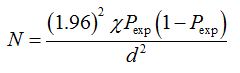
Figure 1: Eggs of Fasciola isolated from cattle faeces by sedimentation technique in the study areas.
| Factor Studied | No of Animals | Prevalence (%) | Chi-square () | P-value | |
| Examined | Positive | ||||
| Study Site | |||||
| Kombolcha | 112 | 24 | 37.50 | 7.974 | P>0.05 |
| Harbu | 143 | 79 | 55.24 | ||
| Kemissie | 125 | 58 | 46.40 | ||
| Age | |||||
| ?1 year | 54 | 15 | 27.77 | 9.888 | 0.007 |
| 1-5 years | 155 | 81 | 52.25 | ||
| ?5 years | 171 | 83 | 48.53 | ||
| Sex | |||||
| Male | 172 | 81 | 47.09 | 0.000 | P>0.05 |
| Female | 208 | 98 | 47.11 | ||
| Total | 380 | 179 | 47.10 | ||
Table 1: The prevalence of Bovine Fasciolosis based on study site, sex and age categories.
Abattoir survey
| Factor Studied | No of Animals | Prevalence (%) | Chi-square () | P-value | |
| Examined | Positive | ||||
| Month | |||||
| December | 117 | 60 | 51.28 | 3.975 | P>0.05 |
| January | 136 | 73 | 53.67 | ||
| February | 127 | 72 | 56.69 | ||
| Total | 380 | 205 | 53.97 | ||
Table 2: Monthly prevalence of Bovine Fasciolosis based on abattoir survey.
| Factor Studied | No of Livers Positive | Prevalence (%) | Chi-square () | P-value | |
| Species of Parasite | |||||
| F. hepatica | 103 | 50.24 | 11.614 | P?0.05 | |
| F. gigantica | 75 | 36.58 | |||
| Mixed infection | 27 | 13.17 | |||
| Total | 205 | 53.97 | |||
Table 3: Prevalence of Bovine Fasciolosis based on species of parasite at abattoir survey.
Financial loss analysis
• Annual estimated value of condemned liver = NAL x CL x %condemnation, where NAL = Average number of cattle slaughtered at Kombolcha ELFORA abattoir; CL = Mean cost of one liver in Kombolcha town; % cond. = Percentage of liver condemned due to fasciolosis
= 4,435 x 14 x 53.97%
= 33,509.97 Ethiopian Birr (US$ 1,435.73)
• Indirect annual due to reduction = NAL x CL x PA x Prev. in meat production, where: NAL = Average number of cattle slaughtered in the meat factory per year; CL = Carcass weight loss in individual animals due to Fasciolosis; PA = Average market price of one kilogram of beef in Kombolcha town; Prev. = Prevalence of Fasciolosis in the meat factory
= 4,435 x (126 x 10%) x 52 x 53.97%
= 1,568,266.74 Ethiopian Birr (US$ 67,192.23)
The total annual financial loss due to fasciolosis in the meat factory of the study area is therefore; 1,601,776.71 Ethiopian Birr (US$ 68,627.97).
DISCUSSION
The results of faecal examination in males and females were 47.09% and 47.11%, respectively. There was no significance difference (p>0.05) in susceptibility to fasciolosis suggesting sex seems to have no impact on the infection rate of fasciolosis. Both male and female animals were equally exposed to the disease. Similar results that support the present findings were reported by Dagne and Rahemato [24,25]. On the contrary, Balock and Arthur revealed high prevalence in males than females [26]. This might probably related to the management system with longer exposure of male outdoor while females are kept in-door during pregnancy and lactation. This study indicated a prevalence of 27.77%, 52.25% and 48.53% among age groups <1 year, 1-5 years and >5 years, respectively. A significant variation (p<0.05) was recorded in the infection rate between different age groups, with higher in 1-5 years age group while the lower was observed in age group of <1 year. This finding agreed with the work of Solomon and Abebe [27]. This may be attributed to the fact that young animals were not often driven far with older age groups to grazing and watering points. They were mostly kept at a nearby village where the sources of feeding sites are not contaminated. This practice naturally reduces the chance of exposure in this age class. The more the age of the young increases, the possibility of moving towards new environment happens, which leads to an exposure with Fasciola contaminated pasture lands and water points. According to Rahemato, similar results indicated inverse correlation of prevalence and age of cattle in different parts of Ethiopia [25].
The result of Kombolcha ELFORA abattoir survey revealed the prevalence of 53.97% Bovine Fasciolosis. This result seems to be lower compared to the results of previous reports in other parts of the country; such as 86% in Keffa and 70% in Illubabur Administrative Region, 80% in and around Debre Berhan and 82.5% in Western Shoa, 71% at Addis Ababa abattoir, 75% at Gondar municipal Industrial abattoir and 77.8% at Dembidolo abattoir [4,5,24,27-29]. This variation in the prevalence probably due to difference in climatic condition (altitude, rain fall, temperature) of the area, management system of animals and availability of veterinary services and drug usage.
However, the result observed at Kombolcha ELFORA abattoir is relatively close to those obtained at Debre Zeit abattoir 49%, at Jimma municipal abattoir, 47% and at Gondar municipal abattoir, 49% [3,22,30]. All the Fasciola species observed in the infected livers were identified as F. hepatica and F. gigantica. The occurrence of F. hepatica, F. gigantica and mixed infection by both species at the abattoir is 50.24%, 36.58% and 13.17%, respectively. This result was very close to Abegaz where 50.07% of F. hepatica, 39.63% F. gigantica and 10.29% mixed infection at Kombolcha ELFORA abattoir [31]. On the other hand, result of Mitiku at Bedele municipal abattoir was higher for F. hepatica (64.5%) and lower for F. gigantic (24.8%) when compared to the present study result [32]. Mixed infection by both species of Fasciolamight be due to cattle for slaughter normally comes from different marketing areas of the region having different weather conditions and altitude known to be suitable for the existence of both species of Fasciola and intermediate hosts. The study conducted by Graber indicated the Ethiopian F. hepatica found in areas situated over 1800-2000m asl, F. gigantica up to 1200m asl and mixed infection with both species in areas between 1200-1800m asl [2].
Although it was difficult to evaluate the actual financial loss incurred due to individual parasitic diseases, because of the occurrence of polyparasitism in the natural case, financial loss analysis due to Fasciolosis was made at Kombolcha ELFORA abattoir. This was done using the number of livers condemned per year and carcass weight loss which is indirectly associated with liver pathology. In doing so a sum of money amounting 33,509.97 Ethiopian Birr (US$ 1,435.73) was lost due to liver condemnation and 1,568,226.74 Ethiopian Birr (US$ 67,192.23) as a result of reduction in meat production with a total loss of 1,601,776.71 Ethiopian Birr (US$ 68,627.97) annually due to Fasciolosis. Financial loss analysis reported from other part of the country include Mulugeta in Kombolcha (287,911.32 Ethiopian Birr), Wondoson in Arsi (159,704.00 Ethiopian Birr) and Mitiku in Jimma per annum [32-34]. These results showed that fasciolosis cause significant lose in different parts of Ethiopia at large. Considering the prevalence of the disease and its economic significance in different parts of the country, one can strongly conclude that Fasciolosis is one of the most important livestock parasitic diseases which impose huge carcass condemnation.
CONCLUSION
ACKNOWLEDGEMENT
REFERENCES
- Sintayehu (1998) VM Research Proposal on Liver Fluke of Cattle in and Around Sebeta. Field and abattoir survey, Addis Ababa University, Ethiopia. Pg no: 1-41.
- Graber M (1975) Helminthes and Helminthiasis of Domestic and Wild Animals of Ethiopia. Bulletin of Animal Health and Production in Africa 23: 57086.
- Fiseha, Yilema J (1983) Economic Importance of bovine Fasciolosis: An assessment trail at Debre Zeit abattoirs. 2nd Students scientific journal, Addis Ababa University, Ethiopia. Pg no: 28.
- Getachew T (1984) A survey of fasciolosis in cattle, sheep and goats slaughtered at Addis Ababa abattoir. IPP research report, Addis Ababa University, Ethiopia. Pg no: 10-11.
- Bahru G, Ephraim M (1979) A Preliminary Survey of Bovine Fascioliasis in Ethiopia. Ethiopian Journal of Agricultural Sciences.
- Urqhuart GM, Armour J, Duncan JJ, Dunn AM, Jennings FW (1996) Veterinary Parasitology (2ndedn). Wiley, Hoboken, New Jersey, USA. Pg no: 307.
- Stuart MT (1997) Fluke infection in ruminants, lung worm infection: The Merck veterinary manual (8thedn).
- Gaafar SM, Howard WE, Marsh RE (1985) Parasites, pests, and predators. Elsevier Scientific Pub. Co., Amsterdam, Netherlands. Pg no: 575.
- Hillyer GV, Apt W (1997) Food-borne trematode infections in the Americas.Trends in Parasitology 13: 87-88.
- WHO (1995) Control of foodborne trematode infections. WHO, Geneva, Switzerland.
- Fabiyi JP (1987) Production losses and control of helminths in ruminants of tropical regions. Int J Parasitol 17: 435-442.
- Schillhorn Van veen TW (1980) Fascioliasis (Fasciola gigantica) in West Africa: a review. Veterinary Bulletin 50: 520-533.
- Njau BC, Scholtens RG (1991) The Role of Traditionally Harvested hay in the transmission of Ovine Fasciolosis in the Ethiopian highlands. Vet Res Commun 15: 369-372.
- Hall MTB (1988) Disease and Parasite of livestock in the tropics. Langman, London, England. Pg no: 328.
- Soulsby EJL (1982) Helminths, Arthropods and Protozoa of Domesticated Animals (7thedn). Lea & Febiger, Philadelphia, Pennsylvania, USA. Pg no: 809.
- Amhara Regional Bureau of Agriculture (2006) Annual regional agricultural report, Bahir Dar, Ethiopia.
- Central Statistical Authority (2003) Ethiopian Agricultural Sample Enumeration, 2001/02 (1994 E.C.): Statistical report on livestock and farm implements. Central Statistical Authority, Ethiopia.
- Frandson RD (1974) Anatomy and Physiology of Farm Animals (2ndedn). Philadelphia Lea & Febiger, Philadelphia, Pennsylvania, USA. Pg no: 445-447.
- ILCA (1991) ILCA ... Annual Report and Programme Highlights. ILCA, Addis Ababa, Ethiopia.
- Ogunrinade A, Adegoke GO (1982) Bovine fascioliasis in Nigeria--intercurrent parasitic and bacterial infections. Trop Anim Health Prod 14: 121-125.
- Thrusfield M (1997) ELST - Veterinary Epidemiology (2ndedn). John Wiley & Sons Limited, New York City, USA. Pg no: 504.
- Yehenew M (1985) Prevalence of Fasciolosis in Gondar clinic and Around Lake Tana. DVM Thesis, FVM, Addis Ababa University, Debre Zeit, Ethiopia. Pg no: 1-42.
- Fekadu R (1988) A preliminary Survey on bovine Fasciolosis around Bahir Dar. DVM Thesis, FVM, Addis Ababa University, Debre Zeit, Ethiopia. Pg no: 1-45.
- Dagne M (1994) Survey on Prevalence and Economic Significance of Bovine Fasciolosis in Debre Berhan region. DVM Thesis, FVM, Addis Ababa University, Debre Zeit, Ethiopia. Pg no: 1-44.
- Rahemato A (1992) Fasciolosis: Clinical occurrence, coprological, abattoir and sna il survey in and around Wolisso. DVM Thesis, FVM, Addis Ababa University, Debre Zeit, Ethiopia. Pg no: 1-46.
- Balock FC, Arthur RJ (1985) A survey of fascioliasis in beef cattle killed at abattoirs in southern Queensland. Australian Veterinary Journal 62: 324-326.
- Solomon A, Abebe M (2007) Prevalence and Economic Significance of Bovine Fasciolosis at Nekemt. DVM Thesis, Addis Ababa University, Debre Zeit, Ethiopia. Pg no: 1-37.
- Yadeta B (1994) Epidemiology of Bovine and Ovine Fasciolosis and Distribution of its snail intermediate host in Western Shoa. DVM Thesis, FVM, Addis Ababa University, Debre Zeit, Ethiopia. Pg no: 1-35.
- Roman T (1987) Study on economic significance of bovine fasciolosis and hydatidosis at Gondar abattoir. DVM Thesis, FVM, Addis Ababa University, Debre Zeit, Ethiopia. Pg no: 1-30.
- Zewdu B (1991) Prevalence and economic analysis of liver fluke infestation in cattle slaughtered at Jimma municipal abattoir. DVM Thesis, Addis Ababa University, Debre Zeit, Ethiopia. Pg no: 1-37.
- Abegaz S (2001) Epidemiology, Control and Economic Importance of Bovine Fasciolosis in Cheffa Valley of the Eastern Amhara Region. Regional Veterinary, Ethiopia. Pg no: 1-43.
- Mitiku W (2009) Prevalence and Economic significance of Bovine Fasciolosis at Bedele Municipal Abattoir. DVM Thesis, Jimma University, Ethiopia. Pg No: 1-29.
- Mulugeta T (1993) Prevalence and Economic Significance of Bovine Fasciolosis at Sopral Kombolcha Meat Factory. FVM, Addis Ababa University, Debre Zeit, Ethiopia. Pg no: 1-38.
- Wondoson A (1990) Prevalence of Bovine Fasciolosis in Arsi Administrative Region. DVM Thesis, Addis Ababa University, Debre Zeit, Ethiopia. Pg no: 1-32.
Citation: Ayelign M, Alemneh T (2017) Study on Prevalence and Economic Importance of Bovine Fasciolosis in Three Districts of North-East Amhara Region, Ethiopia. J Infect Non Infect Dis 3: 024.
Copyright: © 2017 Tewodros Alemneh, et al. This is an open-access article distributed under the terms of the Creative Commons Attribution License, which permits unrestricted use, distribution, and reproduction in any medium, provided the original author and source are credited.