Mucinous Appendiceal Neoplasia: Case Report and Literature Review
*Corresponding Author(s):
Rafael Francisco Alves SilvaGraduate Student, Medical School, University Center Of Brasilia (UniCEUB), Brasilia, Brazil
Tel:+55 61999232232,
Email:rafafalves97@gmail.com
Abstract
Introduction
Primary appendececal tumors are rare occurrences, found in less than 2% of appendectomies. Aim of this study is to report a case of acute appendicitis caused by Low Grade Mucinous Neoplasia (LAMN) and to review histopathological classification and treatment in the literature.
Case report
75 years-old, female, complaint of pain in the right iliac fossa for 15 days, associated with nausea and hyporexia for 1 day. At physical examination, diffuse pain at palpation of right hemiabdome. Hemogram showed 15.200 leukocytes, with 4% of rods. Ultrasonography of abdomen without alterations and computed tomography suggestive of appendicitis. During exploratory laparotomy appendix was enlarged, hyperemic with thick walls and hyperemic cecum with signs of ischemia, suggesting a neoplastic process. Opted for right colectomy with primary anastomosis. Patient progresses well with discharge in the 10thpostoperative. Histopathological analysis of low grade mucinous neoplasm in the appendix.
Discussion/conclusion
The classification of appendiceal tumors is controversial; LAMN is a submucosal restricted epithelial tumor with low degree of cellular atypia. Treatment is based on removal of the primary site of injury by appendectomy or colectomy.
INTRODUCTION
Primary appendececal tumors are rare occurrences, found in lessthan 2% of appendectomies [1]. The mucinous appendix neoplasia accounts for 0.2-0.7% of the cases [2]. Diagnosis is usually performed in the sixth decade of life [3]. Clinical manifestations range from absence of symptoms, acute appendicitis, non-spastic abdominal pain and palpable abdominal tumor [2]. The surgical treatment can be performed appendectomy or hemicolectomy, according to the extent of the lesion [4]. The aim of this study is to report a case of acute appendicitisca used by Low Grade Mucinous Neoplasia (LAMN) and to review histopathological classification and treatment in the literature.
CASE REPORT
75 years-old, female, hypertensive and diabetic. He was admitted in the Hospital, complaint of pain in the right iliac fossa for 15 days, associated with nausea and hyporexia for 1 day. Denyvomiting, diarrhea and urinary symptoms. Physical examination showed (HR: 88bpm, SatO2: 98%) semi globous abdomen, abdominal auscultation without alterations, with diffuse pain on right hemiabdomepalpation. Hemogramwith 15,200 leukocytes, 4% of rods.
Ultrasonography of total abdomen performed on the day of care without alterations. Computed tomography showed a distended, thickened appendix with adjacent adipose densification, with no associated perforationor collection signs, suggesting a diagnosis of appendicitis (Figure 1). Anexploratory laparotomy was indicated for possible appendectomy.
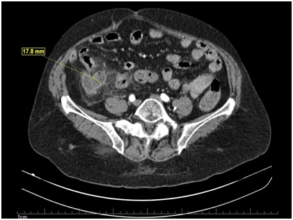 Figure 1: Computed Tomography (CT) of sagittal incidence evidencing a distended appendix, thickened with adjacent adipose densification.
During the intra operative period, thececal appendix was observed, blocked by the largeomentum. The appendix was enlarged, hyperemic with thick walls and hyperemiccecum with signs of ischemia, suggesting a neoplastic process (Figure 2). Absence of extra vasation of fecal or mucinous contents in the peritoneal cavity, without lymph node enlargementor peritoneal or hepatic implants. Right colect omy was chosen, with primary anastomosis. Patient progressed uneventfully, being discharged on the 10thpostoperative day. On the 17th postoperative day, here turned to the hospital with a complaint of surgical wound infection, opted for conservative treatment with antibiotic therapy and wound cleaning.
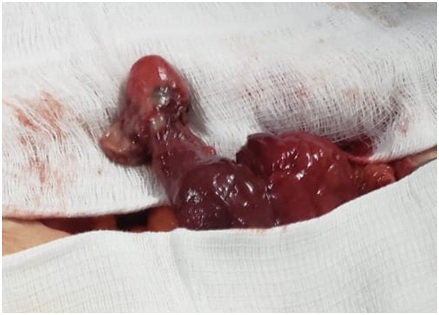 Figure 2: Aspect of the appendix during the intraoperative.
Histopathological analysis of the surgical specimen presentedcecal appendixw ith 5.0x3.0cm. Presence of low grade, well differentiated appendicular mucinous neoplasia with extension in every organ, with tumor free margin and lymph nodes without involvement (Figure 3).
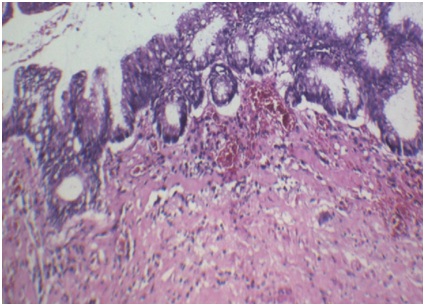 Figure 3: Histopathological analysis of the appendix, H&E stain. Presented dysplastic epithelium, sometimes venous, sometimes tubular, with a presence of mucin in the cytoplasm of the cell.
DISCUSSION
The classification of appendix tumors is controversial. Mucin-producing tumors have epithelial origin, according to WHO classification of 2010, mucin-producing neoplasms were divided into 3 groups: Adenomas, LAMN and adenocarcinoma of the apêndix [1].
Currently, by the 2016 Modified Delphi Consensus Protocol the mucinous neoplasms are classified as: LAMN, high grade mucinous neoplasia, mucinous adenocarcinoma with or without signet cell [5].
LAMN and high-grade mucinous neoplasia are lesions restricted to the submucosa and are differentiated by the level of cellular atypia, although these lesions are not malignant, they may present extra appendicular implantation. Mucinous adenocarcinoma is a malignant lesion, whichhas its own muscular invasion [6]. It isbelievedthat LAMN is the initial neoplastic process, with an intermediate stage marked by high grade atypia followed by mucinous adenocarcinoma. The presence of ringcells is indicative of a worse prognosis [7].
The main complicationo f LMNA is the rupture of the appendix causing pseudomyxoma peritonei, a clinical condition characterized by peritoneal neoplastic implantation with accumulation of ascites and fatal outcome [1,2,4,7,8].
LMNA treatment is based on removal of the primary lesions ite by appendectomy [7]. Colectomy is indicated when the tumor is largerthan 2cm in size or involvement of the pericecal región or signs of malignancy [4]. In case of pseudomyxoma peritonei, surgical cytoreduction and hyperthermic intraperitoneal chemotherapy are always indicated [4,7,8].
CONCLUSION
The LMNA of appendixis a raredisease, usually the diagnosis is made in the intraoperative appendectomy. The histopathological classifications are controversial and the treatment varied, so the surgeon and the pathologist must be attentive to the patient's best conduct and follow-up.
REFERENCES
- Tirumani SH, Fraser-Hill M, Auer R, Shabana W, Walsh C, et al. (2013) Mucinous neoplasms of the appendix: A current comprehensive clinicopathologic and imaging review. Cancer Imaging 13: 14-25.
- Pantiora EV, Massaras D, Koutalas J, Bagiasta A, Kontis EA, et al. (2018) Low-grade appendiceal mucinous neoplasm presenting as adnexal mass: A case report. Cureus 10: 3568.
- Teixeira FJR, Do Couto Netto SD, Akaishi EH, Utiyama EM, Menegozzo CAM, et al. (2017) Acute appendicitis, inflammatory appendiceal mass and the risk of a hidden malignant tumor: A systematic review of the literature. World J Emerg Surg 12: 12.
- Shaib WL, Assi R, Shamseddine A, Alese OB, Staley C 3rd, et al. (2017) Appendiceal mucinous neoplasms: Diagnosis and Management. Oncologist 22: 1107-1116.
- Carr NJ, Cecil TD, Mohamed F, Sobin LH, Sugarbaker PH, et al. (2016) A consensus for classification and pathologic reporting of pseudomyxoma peritonei and associated appendiceal neoplasia: The results of the Peritoneal Surface Oncology Group International (PSOGI) modified delphi process. Am J Surg Pathol 40: 14-26.
- Gundogar O, Kimiloglu E, Komut N, Cin M, Bektas S, et al. (2018) Evaluation of appendiceal mucinous neoplasms with a new classification system and literature review. Turkish J Gastroenterol 29: 533-542.
- Legué LM, Creemers G-J, de Hingh IHJT, Lemmens VEPP, Huysentruyt CJ (2018) Review: Pathology and its clinical relevance of mucinous appendiceal neoplasms and pseudomyxoma peritonei. Clin Colorectal Cancer 1533-0028: 30467-30475.
- Bartlett DJ, Thacker PG, Grotz TE, Graham RP, Fletcher JG, et al. (2019) Mucinous appendiceal neoplasms: Classification, imaging, and HIPEC. Abdominal Radiology Pg no: 1-17.
Citation: Silva RFA, Souza LCA, Alves AR, De Paiva VB, Torres AP, et al. (2019) Mucinous Appendiceal Neoplasia: Case Report and Literature Review. J Surg Curr Trend Innov 3: 012
Copyright: © 2019 Rafael Francisco Alves Silva, et al. This is an open-access article distributed under the terms of the Creative Commons Attribution License, which permits unrestricted use, distribution, and reproduction in any medium, provided the original author and source are credited.