Dynamic Ultrasound for Medial Meniscus Tear (Transverse)
*Corresponding Author(s):
Abdulwahed H AlzaherSonographer, Department Of Physical Therapy, Jubail General Hospital, King Abdullah, Al Jubail, Saudi Arabia
Tel:+966 13 362 2233,
Email:abdu203070@gmail.com
Abstract
Ultrasonography is a significant imaging modality as sonographers use it to examine and diagnose pathology. Ultrasound capture images of internal organs such as bones, joints, blood vessels, and muscles, thereby enabling medical practitioners to diagnose diseases. This report seeks to examine medical meniscal tear (MM) and the potential implications of transverse using ultrasound. The report examines the use of valgus and stress tests in providing a clear and accurate image of the pathologies of the meniscus. Ultrasonography is a useful medical imaging technology as it is cost-efficient, readily available, non-invasive, and reliable. Its broad application limits the use of arthroscopy and MRI. Ultrasound is a critical medical innovation as it supports the evaluation and diagnosis of pathogens in the musculoskeletal system.
INTRODUCTION
Ultrasonography is a useful medical imaging modality as it uses ultrasound to create images of internal body structures such as joints, tendons, internal organs, muscles, and blood vessels. Primarily, ultrasonography helps sonographers view pathologies or determine the source of a disease. Besides, ultrasonography is cost-effective, reliable, and non-invasive. Sonographers also prefer ultrasonography as this medical innovation has a spectrum of clinical applications. Using technologies such as power Doppler sonography and high-resolution probes, ultrasonography performs improved medical imaging as compared to other medical imaging procedures. As such, it helps physicians to create visual representations of joints, ligaments, muscles, and tendons for medical intervention and clinical analysis. Likewise, ultrasonography is a useful medical technology as it helps clinicians to examine and treat musculoskeletal disorders such as meniscus tears. This article demonstrates how dynamic sonography can diagnose musculoskeletal disorders such as medial meniscus tear (Transverse).
ANATOMY
Anatomy is a critical branch of biology as it deals with the study of the structure and parts of organisms. Human anatomy explains how different body parts interact to form a functional unit. Bones are essential components of human anatomy as they facilitate movements. Besides, bones support muscles and tendons. Physical therapists (PTs) use sonography to examine medical meniscal tear. Notably, medial meniscus tear injuries are common as during injury, medial meniscus absorbs significant shock [1].
Additionally, supine valgus and supine Varus scanning helps medical practitioners to examine the tibiofemoral joint (TFJ). This technology uses a transverse axis device to divide the body for proper medical imaging [2]. Using a short axis of the transducer in place of the TFJ line, sonography reveals the minuscular tear with a 30° supine flexionand dynamic movement of the knee. Again, the ultrasound imaging of medial collateral ligament (MCL) reveals two layers of the bone (super facial and deep layer), which are separated by hypoechoic layers. However, using static images, it is challenging to study the anatomy of the meniscus tear and diagnose pathology as this technique does not capture moving images.
CASE REPORT
The case involves a 35 years old patient who plays football. He presents to a health unit with a knee injury, on the right side of the medial. The patient explains that he twisted his knee two weeks ago and experiences pain. The PT uses the McMurray method to examine the injury and notes that there is no swelling on the knee. The ultrasound setting is a health center. The examination takes 10 minutes. The PT uses an ultrasound transducer that receives and transmits echoes to a computer. This device produces sound waves with frequencies that range between 2 and 15 MHz.
PROGNOSIS OF A KNEE INJURY
The prognosis of the knee injury started from the twisting of the leg. The twisting of the knee caused a medial meniscus tear. The injury caused inflammation in the knee as the knee tissues were damaged. As such, the patient experiences pain. However, since the injury was not severe, the injured area does not swell. Treatment for this injury includes pain relievers, which help manage pain. Besides, PT uses physiotherapies to prevent swelling and reddening of the affected area [3].
ULTRASOUND APPEARANCE OF THE CASE
The ultrasound reveals injured ligaments in the right side of the knee. The ligaments have mixed echogenicity. Besides, medical imaging shows meniscal injuries. Again, sonography reveals muscle tears, which cause pain in the knee. The ultrasound also reveals effusion in inflammatory ultrasound appearance of knee injury synovial thickening, and erosion of the articular surface.
MENISCAL TEAR CLASSIFICATION (GRADE I, II, III)
Meniscal tear grade I encompass small focus as the injury is not severe. Ultrasound reveals insignificant tissue damages to the affected region. Grade II of the meniscal tear has a linear area. This injury is not severe and reveals insignificant symptoms. Grade III encompasses a complete meniscal tear. This damage is acute as it represents articular surface damage. Ultrasound shows that the twisted knee is in a static position, as revealed in the following figures 1-3[4].
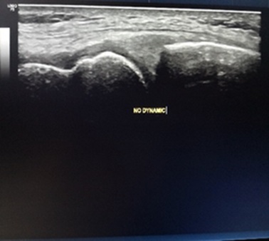 Figure 1: An image of medial meniscus using static image.
However, the image of the medial meniscus tear was clear using a valgus stress test, as revealed below.
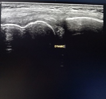 Figure 2: An image of medial meniscus tear using ultrasonography.
Using the dynamic (valgus stress test), the image becomes clear and can be measured as revealed below.
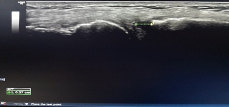 Figure 3: Measurement of the tear (transverse tear) with last dynamic valgus stress.
DISCUSSION
Ultrasound is a useful imaging technique as it detects meniscus pathology. The above case study shows that ultrasound creates clear images that help medical practitioners to treat diseases. However, although ultrasound help to checks and treat meniscus pathology, magnetic resonance imaging (MRI) is preferred as this medical imaging use modernized scanners that use computers, radio waves, and magnetic fields to generate images of internal body organs [4]. Therefore, if a patient complains of knee trauma, we recommend the use of ultrasound screening tools to check meniscal injuries. Besides, we recommend physical examinations using the McMurray technique to check if the condition of the knee. Again, we would prefer the use of X-rays, CT scan, and nuclear medicine imaging. If these methods fail to offer reliable medical data, we would recommend MRI as this technique creates images of physiological processes and anatomy. The following are images of the twisted knee created by MRI(Figures 4,5)[3].
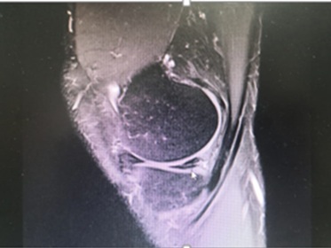 Figure 4: Image of an injured knee.
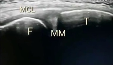 Figure 5: An image of an MCL created by MRI technique.
CONCLUSION
Ultrasonography uses ultrasound technologies to capture images of joints, muscles, and blood vessels. The technology is useful as it helps medical practitioners detect pathologies. For example, it takes images of a medial meniscus tear. Therefore, it allows physicians to determine optimal medication methods for the patient. However, MRI is preferred to ultrasonography as it uses modern techniques to take images of body organs.
REFERENCES
- Bolog NV, Andreisek G (2016) Reporting knee meniscal tears: technical aspects, typical pitfalls and how to avoid them. Insights Imaging 7: 385-398.
- Lecouvet F, Van Haver T, Acid S, Perlepe V, Kirchgesner T, et al. (2018) Magnetic resonance imaging (MRI) of the knee: Identification of difficult-to-diagnose meniscal lesions. Diagn Interv Imaging 99: 55-64.
- Ahmed AF, Azeem AA, Eladawy A, Abdeen M (2017) MRI as an accurate tool for the diagnosis and characterization of different knee joint meniscal injuries. Egypt J Radiol Nucl Med 48: 953-960.
- Lefevre N, Naouri JF, Herman S, Gerometta A, Klouche S, et al. (2016) A Current Review of the Meniscus Imaging: Proposition of a Useful Tool for Its Radiologic Analysis. Radiol Res Pract 2016:8329296.
Citation: Alzaher AH, Alnizi RL (2019) Dynamic Ultrasound for Medial Meniscus Tear (Transverse). J Nucl Med Radiol Radiat Ther 4: 015.
Copyright: © 2019 Abdulwahed H Alzaher, et al. This is an open-access article distributed under the terms of the Creative Commons Attribution License, which permits unrestricted use, distribution, and reproduction in any medium, provided the original author and source are credited.