Conjunctival Melanoma and Role of Immunohistochemical Markers protein S100, HMB-45 and Melan A in Tumor Staging: Case Report and Literature Review
*Corresponding Author(s):
Luiz Carlos De Araújo SouzaResearcher Of The Department Of Cytopathology And Pathological Anatomy Of The Base Institute Of The , St. Hospital Medical South - Asa Sul, Brasilia, Brazil
Tel:+55 61999232232,
Email:luiz_carlos5@hotmail.com
Abstract
We describe the case of a 84-year-old male with hyperdensitive and heterogeneous expansive lesion on the right eyelid suggestive of Conjunctival Melanoma (CM). They performed orbital exenteration with graft of the abdominal wall in the orbital cavity. Surgical specimen analysis with S100, HMB-45 and Melan A positive showed a CM with invasion of the right eyelid, intact optic nerve, clean surgical margins and absence of angiolymphatic invasion.
Keywords
INTRODUCTION
Conjunctival Melanoma (CM), a malignancy derived from melanocytes located in the basal layer of the conjunctival epithelium, is the second most frequent lesion of the conjunctiva, after squamous cell carcinoma. It corresponds to nearly 2% of ocular melanomas and less than 1% of all malignant tumors of the eye, being responsible for 5-7% of all primary ocular melanomas [1-3]. Recently, there has been a significant increment in the incidence of CM in men, remaining unaltered in women [4,5].
The prognostic factors known for CM include distinct tumor sites, such as non-bulbar conjunctiva (tarsal or fornix conjunctiva and caruncle), increase in tumor width, local tumor recurrence, high mitotic rate, epithelioid histiocytes, multifocal tumors, myxoid cells, depth of > 4 mm, scleral extension, advanced staging of lymph node metastasis, and incomplete surgical excision [6-9]. Therefore, Immune Histo Chemical (IHC) analyses may significantly assist in the differential diagnosis.
CASE REPORT
An 84-year-old male patient, complaining of an expansive mass in right eye occupying the orbit and ipsilateral amaurosis. A non-contrast computed tomography of orbits identified an expansive lesion in right eyelid spontaneously hyperdense, heterogeneous, with no well defined cleavage plane in relation to the bulbus oculi, measuring 3.4x3.0x1.7 cm (9 cm3) and presenting a depression in its anteroinferior aspect, possibly representing an area of ulceration; optic nerve-sheath complex with contour and thickness both preserved; intraconal and extraconal compartments preserved bilaterally; left lacrimal gland topic and with normal dimensions; skeletal structure intact.
A potentially malignant lesion was suspected, hence the indication for right orbital exenteration with a transtarsal access to the orbital cavity, associated with a skin graft from abdominal region.
The anatomopathological macroscopic analysis of the surgical specimen evinced the following: Content of right orbit exenteration composed of bulbus oculi and eyelid; bulbus oculi measuring 2.3x2.3 cm of length, exhibiting a white sclera, a segment of optic nerve measuring 0.5x0.3 cm posteriorly, and a darkly pigmented tumor (4.5x3.5x2.5 cm) that expands through the anterior aspect of the bulbus oculi, compromising the eyelid; cross sections showed a darkly pigmented tumor that does not invade the cavity of the bulbus oculi. The H.E technique demonstrated the presence of atypical cells rich in melanocytic pigment ascending through the epithelium in several sites of conjunctiva, compatible with malignant melanoma, associated with inflammatory infiltrate and central zone of neoplasm exhibiting enlarged epithelioid and fusiform atypical cells with abundant cytoplasm containing melanocytic granules and oval nuclei (Figure 1). IHC was positive for the biomarkers: protein S100, HMB-45 and Melan A (MART-1) (Figures 2, 3 and 4).
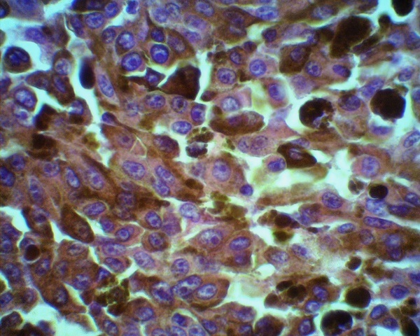 Figure 1: Neoplasm central zone exhibiting enlarged epithelioid and fusiform atypical cells with abundant cytoplasm containing melanocytic granules and oval nuclei widened in size with prominent nuclear membrane and evident peripheral chromatin and eosinophilic macronuclei (HE-400x).
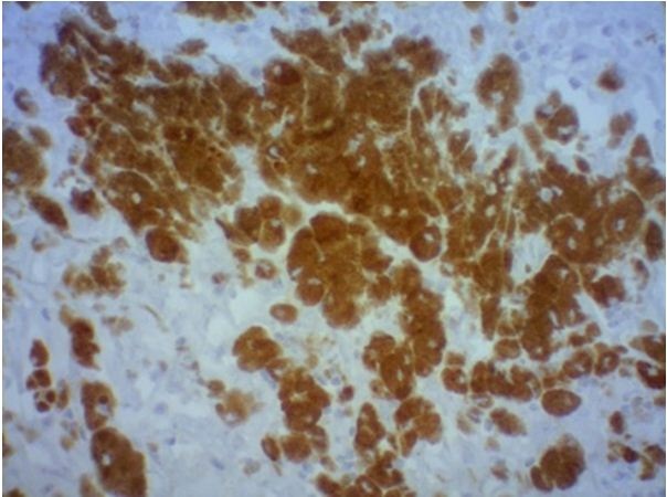 Figure 2: Immunohistochemistry demonstrating positivity for the protein S 100 marker.
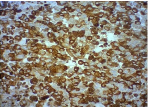 Figure 3:Immunohistochemistry demonstrating positivity for the HMB-45 marker.
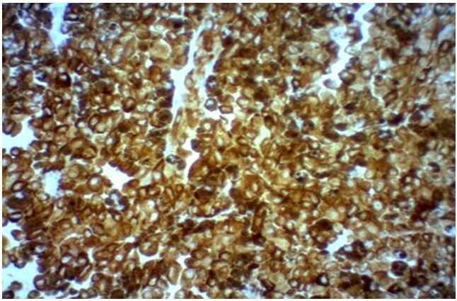 Figure 4:Immunohistochemistry demonstrating positivity for marker Melan A.
Confirming CM of right bulbar of 4.5x3.5x2.5 cm; with tumor extension to conjunctival right eyelid; clear ocular cavity, optic nerve, and surgical margins; and absence of angiolymphatic invasion. The pathological staging was defined as pT3Nx.
DISCUSSION
Protein S100
HMB-45
HMB-45 is therefore very useful and specific for melanocytic tumors, although its sensitivity for melanoma – approximately 70-90% is less than the one of S100. The specimen of the patient in the case being reported was positive for HMB-45 (Figure 3). The concomitant positivity of S100 and HMB-45 increase the specificity of the diagnosis of melanoma, for the marker HMB-45 has high specificity and low sensitivity.
Melan A (MART-1)
In face of being highly sensitive for melanoma, the latter is classified as a useful marker for detecting sites of nodal micrometastases. Melan A marker is both highly useful and sensitive (85 to 97%) in terms of primary tumors, being its sensitivity 57-92% for melanoma with metastasis, and a specificity of 95 to 100%. In contrast to S100, Melan A is not expressed in dendritic cells of lymph nodes. The specimen of the patient in the above case was positive for Melan A (Table 1 and Figure 4). Although Melan A is useful for the detection of nodal micrometastases and considering that the patient of the case discussed presented a positive specimen for the marker, there were no notable signs of metastasis in the ocular cavity, optic nerve, and surgical margins, and angiolymphatic invasion was absent [10]. The concomitant positivity for markers S100, HMB-45, and MART-1 more firmly corroborates the diagnosis of CM.
|
Biomarkers |
Result |
|
Protein S100 |
Positive |
|
HMB-45 |
Positive |
|
Melan A (MART-1) |
Positive |
Table 1: IHC biomarkers stained in the conjunctival melanoma.
CONCLUSION
Despite the discovery of new techniques for screening and diagnosis, CM is yet a rare tumor and presents aggressive characteristics prone to high metastatic potential and mortality. The serial screening of atypical nevi in an early stage is the most effective method of detecting clinical alterations suggestive of malignancy.
METHODOLOGY
Our literature review used articles surveyed in PubMed (NCBI) from 1976 to 2018 using the keywords: Immunohistochemistry; conjunctival melanoma; Protein S100; HMB-45; Melan A (MART-1). For the literature review, the main scientific papers were selected. After the analysis of the main studies, we collected the main histological findings of the markers, sensitivity and specificity of each marker and their clinical and pathological characteristics.
DISCLOSURES AND ETHICS
The case report was submitted to and approved by the National Council of Health - National Commission of Ethics in Research of Brazil - CONEP and the Research Ethics Committee of the Base Hospital Institute of the Federal District-IHBDF.
ACKNOWLEDGMENTS
We would like to thank the staff of the Nucleus of Cytopathology and Anatomy-Pathology of the Federal District Base Hospital Institute (NUCAP-IHBDF) and Laboratory Lamina for the help and the assistance during the elaboration of this paper.
REFERENCES
- Scotto J, Fraumeni JF, Lee JA (1976) Melanomas of the eye and other noncutaneous sites: epidemiologic aspects. J Natl Cancer Inst 56: 489-491.
- Shields CL, Shields JA (2004) Tumors of the conjunctiva and cornea. Surv Ophthalmol 49: 3-24.
- Vajdic CM, Kricker A, Giblin M, McKenzie J, Aitken J, et al. (2003) Incidence of ocular melanoma in Australia from 1990 to 1998. Int J Cancer 105: 117-122.
- Spencer WH (1996) Ophthalmic pathology and the American Academy of Ophthalmology. Ophthalmology 103: 109-117.
- Yu G. Hu D, McCormick S, Finger PT (2003) Conjunctival melanoma: Is it increasing in the United States? Am J Ophthalmol 135: 800-806.
- Edge SB, Byrd DR, Compton CC, et al. eds. Malignant melanoma of the conjunctiva. AJCC Cancer Staging Manual. 7th edn. New York, NY: Springer, 2010:539-46.
- Tuomaala S, Toivonen P, Al-Jamal R, Kivelä T (2007) Prognostic significance of histopathology of primary conjunctival melanoma in Caucasians. Curr Eye Res 32: 939-952.
- Vang R, Kempson RL (2002) Perivascular epithelioid cell tumor ('PEComa') of the uterus: a subset of HMB-45-positive epithelioid mesenchymal neoplasms with an uncertain relationship to pure smooth muscle tumors. Am J Surg Pathol 26: 1-13.
- Werschnik C, Lommatzsch PK (2002) Long-term follow-up of patients with conjunctival melanoma. Am J Clin Oncol 25: 248-255.
- Ordóñez, NG (2014) Value of melanocytic-associated immunohistochemical markers in the diagnosis of malignant melanoma: A review and update. Hum Pathol 45: 191-205.
Citation: Souza LCA, Coutinho SLBM, dos Santos Medeiros SM, De Figueiredo Cavalcanti HO, De Miranda RL, et al. (2019) Conjunctival Melanoma and Role of Immunohistochemical Markers protein S100, HMB-45 and Melan A in Tumor Staging: Case Report and Literature Review. J Surg Curr Trend Innov 3: 011.
Copyright: © 2019 Luiz Carlos de Araújo Souza, et al. This is an open-access article distributed under the terms of the Creative Commons Attribution License, which permits unrestricted use, distribution, and reproduction in any medium, provided the original author and source are credited.