Journal of Otolaryngology Head & Neck Surgery Category: Clinical Type: Case Report

A Case of IgG4-Related Mikulicz’s Disease with Chronic Rhinosinusitis and Elevated Levels of IgG4-and IgE-Immune Complexes

Yasushi Ota1*, Mutsunori Fujiwara2, Hirabayashi Yoji3, Rumi Ota4 and Mitsuya Suzuki1
1 Department of otorhinolaryngology, Toho University, Tokyo, Japan
2 Department of pathology, Nissan Tamagawa Hospital, Tokyo, Japan
3 Clinical laboratory testing division, Toho University Sakura Medical Center, Chiba, Japan
4 Okamura otorhinolaryngology clinic, Hiroshima, Japan

*Corresponding Author(s):
Yasushi Ota
Department Of Otorhinolaryngology, Toho University, Tokyo, Japan
Tel:+81 434628811,
Email:yasushiota5610@yahoo.co.jp

Received Date: Feb 22, 2018
Accepted Date: Apr 14, 2018
Published Date: Apr 27, 2018

Abstract

We report a case of male IgG4-related Mikulicz’s disease with chronic rhinosinusitis with high serum IgG4-and IgE-Immune Complex (IC). Untreated serum IgG4 was 3,110 mg/dL, while Polyethylene Glycol (PEG)-treated serum IgG4-IC was 1,460mg/dL. Both were markedly elevated (normal IgG4 range <105 mg/dL). Serum IgE and PEG-treated serum IgE-IC was also elevated (normal IgE range <170 IU/mL) at 376 IU/mL and 237 IU/mL, respectively. IgE PEG Precipitated (PP) index was higher than normal (<20%) at 63%. We speculate that serum IgG4 and IgE represented autoantibodies for a component of the paranasal sinus mucosa and submandibular gland tissue.

Keywords

Autoantibody; PEG; Submandibular gland

INTRODUCTION

Mikulicz’s Disease (MD) refers to bilateral and symmetrical swelling of the lacrimal, parotid, and submandibular glands and is based on histological similarities reported by Morgan et al. [1], in 1953. Several recent reports from Japan have revealed that MD is associated with elevated serum IgG4 levels and prominent infiltration of IgG4-positive plasmacytes [2,3]; these findings have resulted in the recognition of MD as a singular systemic IgG4-related plasmacytic disease [4]. Although recent reports have documented cases of refractory Chronic Rhinosinusitis (CRS) accompanied by IgG4 infiltration (IgG4-CRS) [5-8], the mechanism underlying IgG4 infiltration in patients with CRS remains unknown.
 
We here in report a case of MD with CRS in which antigen-specific IgG4 and IgE antibodies were measured in serum samples using an AlaSTAT 3gAllergy assay (Siemens Healthcare Diagnostics AG, Erlangen, Germany). In this case, gel filtration and Polyethylene Glycol (PEG) precipitation were used to measure levels of immune complexes of IgG4 (IgG4-IC) and IgE (IgE-IC) in the patient’s serum. We also discuss the role of IgG4 and IgE in MD with CRS.

CASE REPORT

This case was a 42-year-old male patient who visited our hospital for swelling on both sides of the neck and severe bilateral nasal obstruction. Initial medical examination at our hospital revealed polyps in both nasal cavities. Computed Tomography (CT) of the paranasal sinuses and neck revealed bilateral sinusitis, lacrimal gland swelling and bilateral submandibular glands swelling (Figure 1). The patient has asthma and his white blood cell count and eosinophil proportion were 7180/μL and 16.5%, respectively.


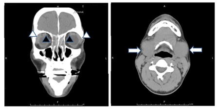
Figure 1: Computed Tomography (CT) of the paranasal sinuses and neck revealed bilateral sinusitis (black arrow heads), lacrimal gland swelling (white arrow heads) and bilateral submandibular glands (white arrows).
 
Endonasal Sinus Surgery (ESS) and biopsy of the left side of his submandibular gland under local anesthesia was performed in November 2013. We diagnosed the patient with IgG4-related MD with CRS and referred him to an autoimmune disease specialist. Below, we detail the histopathological findings of the paranasal mucosa and submandibular gland in this case.

HISTOPATHOLOGICAL FINDINGS

Most of the lymphocytes and plasma cells that had invaded the paranasal lamina propria, as well as the epithelial cells in the parasinus mucosa and the lymphocytes and plasma cells that had invaded the submandibular gland were strongly positive for anti-IgG4 antibody in this case. Vessels and fibrosis tissues were strongly positive for anti-IgG4 antibody. The ratio of IgG4-positive plasma cells to IgG-positive plasma cells was almost 100%. Some mast cells and some of the stroma in the paranasal lamina propria and submandibular gland were positive with anti-IgE antibody (Figure 2).


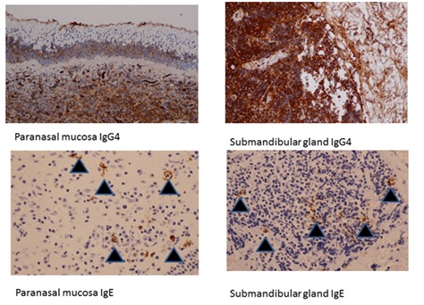
Figure 2: Histopathological findings revealed that most lymphocytes and plasma cells that had invaded the paranasal lamina propria, as well as epithelial cells in the parasinus mucosa and lymphocytes and plasma cells that had invaded the submandibular gland were strongly positive for anti-IgG4 antibody. Some mast cells and some of the stroma in the paranasal lamina propria and submandibular gland were positive for anti-IgE antibody (black arrows).

MEASUREMENT OF ANTIGEN-SPECIFIC IGG4 AND IGE ANTIBODIES IN THE SERUM

The levels of antigen-specific IgG4 and IgE antibodies in this case are shown in table 1. The sum of the antigen-specific IgG4 antibodies was much lower than the total serum IgG4 level and the same was true for IgE (Table 1). IgG4 and IgE measurement were done by the AlaStat 3gAllergy test.

  sIgG4 (mg/dL) sIgE (Iµ/mL)
Total 3110 420
Allergen
Dermatophagoides pteronyssinus 0.6282 <0.10
Dermatophagoide farinae 0.8504 <0.10
House dust-Japan 0.68 <0.10
Japanese Cedar 0.4749 0.479
Dog Dander 0.4529 <0.10
Aspergillus fumigatus 0.5092 <0.10
Mucor racemosus 0.4332 <0.10
Candida albicans 0.5558 <0.10
Alternaria tenuis 0.4288 <0.10
Malassezia furfur (pityrosporum orbiculare) 0.5514 <0.10
Enterotoxin A (S.aureus) 0.8355 <0.10
Enterotoxin B (S.aureus) 0.9488 <0.10
Cockroach 0.8575 <0.10
Table 1: Measurement of antigen-specific IgG4 and IgE antibodies in the serum.

ASSESSMENT OF IMMUNE COMPLEXES IN THE SERUM

Using blood collected from the patient upon his 2013 admission, we used 4% PEG precipitation to measure the levels of circulating IgG4-IC and IgE-IC.
 
Each serum sample was centrifuged at 1000 × g at 4°C for 15 min. Synthetic PEG (Wako Pure Chemical Industries, Ltd.) with a mean molecular mass of 7,500 g/mol was dissolved in 0.01 M phosphate-buffered saline (pH 7.4) at a concentration of 4% (weight/weight). From this stock, 0.1 mL of dissolved PEG was mixed with the serum sample and this mixture was centrifuged at 1000 × g at 4°C for 1 h. The precipitate was dissolved in 0.1 mL phosphate-buffered saline and the IgG4 levels in the precipitate and supernatant (serum) were measured using a paper radioimmunosorbent test. Similar measurements were also performed for IgE. The percentage of IgE in the precipitate vs. that in the serum was calculated and defined as the PEG Precipitated (PP) index.
 
The patient’s level of IgG4 in the untreated serum was 3,110 mg/dL, while the level of IgG4-IC in the PEG-treated serum was 1,460 mg/dL. These IgG4 values were markedly elevated (normal range
 
We measured levels of IgG4, IgE, IgG4-IC and IgE-IC in the sera of two allergic sinusitis cases without MD as controls. Levels of IgG4 were 20.9 and 52.6 mg/dL and those of IgG4-IC were 5.5 and 7.2 mg/dL. Levels of IgE were 1,880 and 1,960 IU/mL and those of IgE-IC were 160 and 122 IU/mL. The IgE PP indexes were 8.5% and 6.2% (Table 2). In IgE, PPI is defined, but in IgG4, IgG, PPI have not defined.

  IgG4 IgE PP Index
PEG-Treated Untreated PEG-Treated Untreated
Serum mg/dL Serum mg/dL Serum mg/dL Serum mg/dL %
Present case 1,460 3,110 237 376 63
Control 1 5.5 20.9 160 1880 8.5
Control 2 7.2 52.6 122 1960 6.2
Table 2: Assessment of immune complexes in the serum.

DISCUSSION

The diagnostic criteria for IgG4-related MD are (i) Symmetrical swelling of at least two pairs of lachrymal, parotid, or submandibular glands for at least 3 months and (ii) Elevated serum IgG4 (>135 mg/dL) or (iii) Histopathological features including lymphocyte and IgG4+ plasma cell infiltration (IgG4+ plasma cells/IgG+ plasma cells >50%) with typical tissue fibrosis or sclerosis [9]. This case fulfilled all three diagnostic criteria and thus a diagnosis of IgG4-related MD was made. The permeation of IgG4-positive cells was seen in the paranasal sinus mucosa of this case and thus the patient also had a diagnosis of IgG4-related CRS. These IgG4-positive cells in the paranasal lamina propria exhibited morphology similar to that of the plasma cells that produce IgG4 in the paranasal mucosa, as previously reported [1-4]. Furthermore, some epithelial cells were stained by anti-IgG4 antibodies, suggesting the presence of some antigens specific for IgG4. Accordingly, we speculated that the IgG4 antibody synthesized by plasma cells was deposited in the epithelium in response to these antigens.
The IgE-positive cells found in the paranasal lamina propria exhibited morphology similar to that of mast cells. These IgE-positive cells may be related to eosinophil permeation in the paranasal sinus mucosa.
To analyze the nature of IgG4 and IgE production in our patient, we identified antigen-specific IgG4 and IgE antibodies in the serum and determined their levels, although the values were all extremely low despite high total serum IgG4 and IgE levels. Therefore, we speculate that the IgG4 and IgE antibodies detected in the serum from this case were nonspecific or specific for an unknown antigen.
 
We thought that the lesion with submandibular gland and the paranasal sinus mucosa was almost the same, because the pathology image resembles each other, for example. A large number of fibrosis tissues and the inflammatory cells appearing in both lesions.
 
In the present study, we used the PEG technique to assess serum immune complexes in this case. The patient’s serum levels of IgG4 and IgE were higher than the normal values and his serum levels of IgG4-IC and IgE-IC were likewise higher than those of two control individuals. His IgE PP index was also high (normal PP index <20%). Thus, large amounts of IgG4-IC and IgE-IC are present in this patient’s serum, which is not observed under general allergy or asthmatic conditions.
 
We hypothesize that the IgG4 and IgE in the patient’s serum were autoantibodies for a component of the paranasal sinus mucosa and submandibular gland tissue because (i) Some epithelial cells of the paranasal mucosa were stained by anti-IgG4 antibodies (ii) The levels of antigen-specific IgG4 and IgE antibodies in his serum were all extremely low despite high total serum IgG4 and IgE levels and (iii) Large amounts of IgG4-IC and IgE-IC exist the patient’s serum. We therefore believe that these autoantibodies caused chronic inflammation of the paranasal sinus and submandibular gland in this case.
 
The IgE autoantibody may cause the protracted type I allergy. It is thought that the IgG4 autoantibody causes intractable inflammation, but the details are unclear.
 
We previously reported a case of female Eosinophilic Granulomatosis with Polyangitis (EGPA) where we used PEG precipitation to evaluate the patient’s levels of IgE-IC, and we speculated that circulating IgE-IC was formed by anti-neutrophil IgE autoantibodies [10]. We herein report a case of male IgG4-related MD with CRS with high serum levels of IgG4-IC and IgE-IC. We propose that the IgG4 and IgE in his serum were autoantibodies for a component of the paranasal sinus mucosa and submandibular gland tissue.

REFERENCES

  1. Morgan WS, Castleman BA (1953) A clinicopathologic study of Mikulicz’s disease. Am J Pathol 29: 471-503.
  2. Yamamoto M, Takahashi H, Sugai S, Imai K (2005) Clinical and pathological characteristics of Mikulicz’s disease (IgG4 related plasmacytic exocrinopathy). Autoimmun Rev 4: 195-200.
  3. Takano K, Yamamoto M, Takahashi H, Shinomura Y, Imai K, et al. (2010) Clinicopathologic similarities between Mikulicz disease and Küttner tumor. Am J Otolaryngol 31: 429-434.
  4. Himi T, Takano K, Yamamoto M, Naishiro Y, Takahashi H (2012) A novel concept of Mikulicz’s disease as IgG4-related disease. Auris Nasus Larynx 39: 9-17.
  5. Takano K, Abe A, Yajima R, Kakuki T, Jitsukawa S, et al. (2015) Clinical evaluation of sinonasal lesions in patients with immunoglobulin G4-related disease. Ann Otol Rhinol Laryngol 124: 965-971.
  6. Inoue A, Wada K, Matsuura K, Osafune H , Ida Y, et al. (2016) IgG4-related disease in the sinonasal cavity accompanied by intranasal structure loss. Auris Nasus Larynx 43: 100-104.
  7. Lee YS, Cho HJ, Yoo HS, Shin YS, Park HS (2014) A case of IgG4-related disease with bronchial asthma and chronic rhinosinusitis in Korea. J Korean Med Sci 29: 599-603.
  8. Ohno K, Matsuda Y, Arai T, Kimura Y (2015) Nasal manifestations of IgG4-related disease: A report of two cases. Auris Nasus Larynx 42: 483-487.
  9. Umehara H, Okazaki K, Masaki Y, Kawano M, Yamamoto M, et al. (2012) Anovel clinical entity, IgG4-Related Disease (IgG4RD): General concept and details. Mod Rheumatol 22: 1-14.
  10. Ota Y, Fujiwara M, Hirabayashi Y, Kumasaka T, Takemura T, et al. (2015) Eosinophilic granulomatosis with polyangiitis demonstrating IgE-immune complexes and the possible involvement of IgE autoantibodies. Intern Med 54: 2687-2691.

Citation: Ota Y, Fujiwara M, Yoji H, Ota R, Suzuki M (2018) A Case of IgG4-Related Mikulicz’s Disease with Chronic Rhinosinusitis and Elevated Levels of IgG4-and IgE-Immune Complexes. J Otolaryng Head Neck Surg 4: 019.

Copyright: © 2018  Yasushi Ota, et al. This is an open-access article distributed under the terms of the Creative Commons Attribution License, which permits unrestricted use, distribution, and reproduction in any medium, provided the original author and source are credited.


Herald Scholarly Open Access is a leading, internationally publishing house in the fields of Sciences. Our mission is to provide an access to knowledge globally.


© 2024, Copyrights Herald Scholarly Open Access. All Rights Reserved!