Correction of Gummy Smile with or without Bone Contouring: A Report of Four Cases
*Corresponding Author(s):
Sudhanshu AgrawalDepartment Of Periodontology, Chandra Dental College & Hospital, Barabanki, India
Tel:+91 9415400591,
Email:drsudhanshu_bds@hotmail.com
Abstract
As quoted in an English book “The Human face “, the first impression of whether one likes or dislikes an individual is often based on what he sees in the face.
Usually, in function of smile, the lips are retracted and elevated to expose a portion of upper incisor teeth .Unlike the case of gummy smile so called “horse smile” there is exposure of all incisors in addition with a large portion of gums above it. A harmonious smile is considered an icon of beauty in modern society. A multifacet scenario including tooth form, position, and gingival tissue levels and lip position determine the smile esthetics. The presence of 3 mm or grater continuous gingival band exposures to natural smile or speech performs the gummy smile ie., (EGD, excessive gingival display).
Gummy smile has been a prevalent esthetic disorder commonly affecting the younger individuals due to various causes such as skeletal, dento -alveolar or soft tissues origin. In due cause for the same, this article enlightens on two different cases in which different approaches for surgical crown lengthening have been perfomed.
Keywords
Gummy smile; Surgical crown lengthening
Introduction
A brilliant, beaming and cheerful smile is an essential feature of beauty to which society gives an increasing notable in the current scenario .The concept of a nice smile basically lean on three anatomic components gums, teeth and lips .Smile line is an imaginary line that follows the lower margin of upper lip and usually has a convex appearance .Excessive exposure of the periodontium characterizes the so-called gingival or gummy smile. This is therefore emphasized that the current esthetic need of the hour standards a dimorphism concept influenced by age and gender, Ergo a healthy and continuous gingival margin display of around 2-3mm during natural smile is considered desirable. EGD include plaque or grug induces gingival enlargement or overgrowth ,short clinical crowns ,Maxillary excess both vertically and anteroposteriorly (Bimaxillary protusion ), hypermobility of upper lip due to hyper function of labial elevator muscles and excessive gingival display associated with altered passive eruption are the major foundation of this condition. In the meantime ,an increasing awareness conceptualizing the awareness regarding beauty and physical appearance has become an impetus for every clinician to evaluate the paramount aspects of patient’s smile and link the dynamic relationship between teeth , gingiva and lips all together while smiling . A part of smile aesthetics is subjective, but most is objective evaluation. The media offers a stereotyped vision of the smile that leads to smile standardization and the new normal, hence an increased patients demand for cosmetic dentistry. EGD can be managed by a variety of procedures that line up as surgical and non surgical methods .The underlying root cause of EGD has the main effect on type of procedure that will be peformed. Non surgical procedures include botulinum toxin type a injection as well as orthodontics while surgical procedures might include lip repositioning or orthognathic surgery following orthodontics .A blend of procedures invoking soft and hard tissues contouring with appropriate prosthetic restoration to alter the shape of the teeth achieves an aesthetic result in an acceptable manner . The rationale for crown lengthing procedures has progressively become more esthetic-driven due to the increasing popularity of smile enhancement therapy (Tables 1&2).
Glimpse Of Literature
|
Authors |
Year |
Acceptable Gingival Exposure |
Having A Gummy Smile |
|
Kokich, et al. |
1999 |
Less than 4mm by laypersons. Less than 2 mm by orthodontists. |
4mm or more by laypersons 2mm or more by orthodontists |
|
Geron, et al. and Atalia, et al. |
2005 |
Until 1mm |
More than 1mm |
|
Van der Geld, et al. |
2011 |
Until 4mm |
More than 4mm |
|
Jannani, et al. |
2014 |
2-3mm |
More then 3mm |
|
Pinto, et al. |
2015 |
1-3mm |
More than 3mm |
|
Bhola, et al. |
2015 |
Until 1mm |
More than 1mm |
|
Pausch, et al. |
2017 |
0-2mm |
More than 2mm |
Table 1: Acceptable gingival exposure and diagnosing of gummy smile in some studies in the literature.
|
Classification |
Characteristics |
Advantages |
Disadvantages |
|
Type I |
Sufficient soft tissues allows gingival exposure of the alveolar crest or violation of biologic width. |
May be performed by the restorative dentist. Provisional restorations of the desired length may be placed immediately |
|
|
Type II |
Sufficient soft tissues allows gingival excision without exposure of the alveolar crest but in violation of biologic width. |
Wil tolerate a temporary violation of the biologic width. Allows staging of the gingivectomy and osseous contouring procedures . Provisional restorations of the desired length may be placed immediately . |
Requires osseous contouring . May require a surgical referral . |
|
Type III |
Gingival excision to the desired clinical crown length will expose the alveolar crest . |
Staging of the procedures and alternative treatment sequence may minimize display of exposed subgingval structures. Provisional restorations of desired length may be placed at second stage gingivectomy |
Requires osseous contouring May require a surgical referral Limited flexibility |
|
Type IV |
Gingival excision will result in adequate band of attached gingiva (insufficient amount of attached gingiva.) |
|
Limited surgical options No flexibility A staged approach is not advantageous. May require a surgical referral. |
Table 2: Proposed Classification System for Aestheic Crown Lengthening Procedures.
Diagnosis
The ladder for establishing a correct diagnosis and a specific plan of treatment is through steps ,firstly by categorising with the most recent proposed classification:
Class I: Very high smile (gummy smile): more than 2 mm of apical display or more than 2 mm of gingival margin to cemento-enamel junction with sound periodontium.
Class II: High smile: between 0mm -2mm of gingival margin or between 0mm and 2mm og gingival margin apical to CEJ with sound periodontium.
Class III: Average smile line:the gingival papillae are visible.
Classs IV: Low smile line: gingival papillae and CEJ are visible.
It is extremely important to note the sexual dimorphism aspect and influence of age on smile .The position and amount of teeth and gingival displayed during smile and speech, height and symmetry of face, thickness ,length and profile of lips ,width of keratinized gingiva, gingival biotype ,facial and lingual bone levels .
Pre-procedural assessment
Prior to developing a suitable treatment plan, it is essential to establish a complete and accurate assessment of the conditions with which the patient presents.
- Reasons for seeking treatment
- Assessment of systemic health and habits
- Height and symmetry of face
- Thickness, length, and profile of lips
- Smile line
- Condition and dimensions of teeth
- Width of keratinized gingiva
- Gingival biotype; and Facial and lingual bone levels, thickness of alveolar
Indications
The indications for crown lengthening are:
Restorative needs
To increase clinical crown height lost due to caries, fracture or wear •
To access subgingival caries •
To produce a „ferrule? for restoration •
To access a perforation in the coronal third of the root
To relocate margins of restorations that are impinging on biological width
Aesthetics
Short teeth
Uneven gingival contour
Gummy smile Contra-indications & Limiting Factors
Inadequate crown to root ratio
Non restorability of caries or root fracture
Esthetic compromise
High furcation Inadequate predictability
Tooth arch relationship inadequacy
Compromise adjacent periodontium or esthetics
Insufficient restorative space
No maintainability
Case 1
Surgical crown lengthening with osseous correction & immediate provisional restoration. A 50-year old male patient with sever attrition in maxillary teeth. Sufficient soft tissue present allowing gingival exposure of the alveolar crest (Figures 1-7).
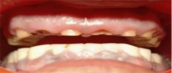 Figure 1: Preoperative Views.
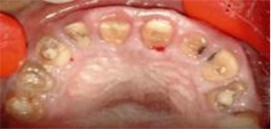 Figure 2: Preoperative Views.
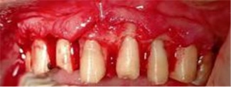 Figure 3: After Flap Reflection Bone Contouring.
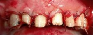 Figure 4: Suturing Was Done.
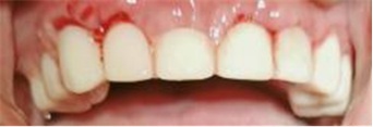 Figure 5: Immediete Provisional Restoration Given.
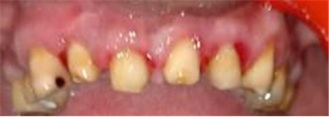 Figure 6: After 3 Month Provisional Prosthesis Removed.
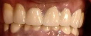 Figure 7: Final Prosthesis was Given.
Case 2
Surgical crown lengthening without osseous correction: A 23-year-old female patient with unaesthetic crowns on the maxillary anterior, and maxillary gingival excess resulting in a gummy smile report (Figures 8-11).
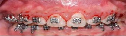 Figure 8: Phase 1 periodontal therapy was performed.
 Figure 9: Bone sounding was done.
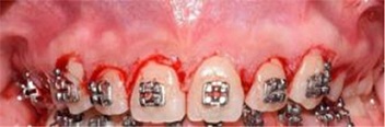 Figure 10: Inverse bevel gingivectomy.
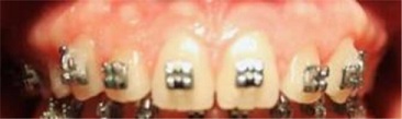 Figure 11: Post opp: Healing after r3 months of healing Discussion.
Case 3
Surgical crown lengthening with osseous correction for gummy smile: A 30-year-old female patient reported to the clinic with a chief complaint of a gummy smile that was not acceptable aesthetically. Adviced surgical crown lengthening involving soft tissue as well as osseous correction (Figures 12-19).
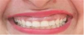
Figure 12: The preoperative view can be seen a gummy smile.
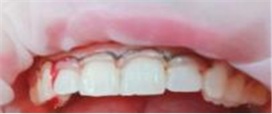 Figure 13: A crevicular incision was made from distal of tooth number, 14 to 24.
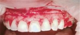 Figure 14: Mucoperiosteal flap was raised.
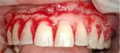 Figure 15: Osseous correction was then performed, keeping in mind the biologic width requirements using a large round diamond bur on an airotor.
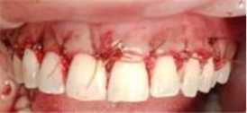 Figure 16: The flap was sutured back in the same position.
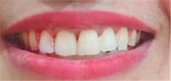 Figure 17: After one weeks of healing and allowing complete shrinkage, gingivectomy was performed with aesthetic contouring.
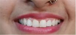 Figure 18: After 1 year of follow up.
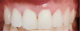 Figure 19: After 1 year of follow up.
Case 4
In 20 years old female patient, crown lengthening with osseous contouring done (Figures 20-24).
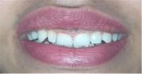 Figure 20: Pre operative view.
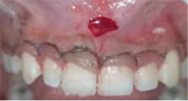 Figure 21: A crevicular incision was made from distal of tooth number, 14 to 24.with frenectomy.
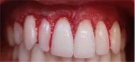 Figure 22: Using a large round diamond bur on an airotor to contour the bone.
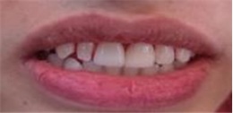 Figure 23: Post operative just after surgery
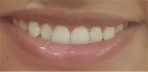 Figure 24: Follow up after 6 months.
There are two aspects to the crown lengthening procedure: Aesthetic and functional. In both cases, the surgical procedure is aimed at re-establishing the biological width, apically, while exposing more tooth structure. Biological width is the sum of the junctional epithelium and supracrestal connective tissue attachment. The average space occupied by the sum of the junctional epithelium and the supracrestal connective tissue fibers was found to be 2.04 mm [2]. Violation of biological width has been associated with gingival inflammation, discomfort, gingival recession, alveolar bone loss, and pocket formation [3].
To have a harmonious and successful long-term restoration, a 3 mm sound supracrestal tooth structure between bone and prosthetic margins, which allows for the reformation of the biological width plus sulcus depth is advocated [4,5]. This can be achieved surgically (crown lengthening), orthodontically (forced eruption), or by a combination of both.
Patients that require aesthetic crown lengthening, however, frequently exhibit a high smile line. As a result, pressure is often placed on the restorative dentist to correct aesthetic deficiencies as early as possible, and maintain certain aesthetic standards throughout the treatment. However, conventional protocols require a waiting period of four to six weeks for sufficient healing of the attachment apparatus, prior to initiating restorative procedures [6]. Hence adequate length of time was allowed for healing in both the cases. The surfaces exposed due to crown lengthening will be displayed through the healing period until the provisional prosthesis can be fabricated or relined. The exposed areas may be limited to cemento-enamel junctions and varying amounts of root surface, but may also include the margins of previous restorations.
Crown lengthening can be limited to the soft tissues when there is enough gingiva coronal to the alveolar bone, allowing for surgical modification of the gingival margins, without the need for osseous recontouring (that is, pseudo pockets in cases of gingival hyperplasia), as seen in case 1, which presented with periodontitis and bone loss. An external or internal bevel gingivectomy (gingivoplasty) is the procedure of choice in these cases.
The biological width has not been compromised, and, as a result, the soft tissue pocket is eliminated and the teeth exposed without the need for osseous resection. Unfortunately, the majority of cases will involve bone recontouring as well as gingival resection to accommodate aesthetics and function. This is a more delicate procedure that requires exposing root surface, positioning gingival margins at the desired height, and apically reestablishing the biological width.
The level of the alveolar crest must be determined prior to any considerations regarding aesthetic crown lengthening. Bone sounding is utilized to determine the thickness of the soft tissue layer and height and thickness of the alveolar bone. Bone sounding assists in determining the level of the alveolar crest, and thus, the need for osseous contouring [7]. The crown-lengthening procedure enables restorative dentists to develop an adequate zone for crown retention without extending the crown margins deep into periodontal tissues. After the procedure, it is customary to wait six to eight weeks before cementing the final restoration. In the aesthetic zone, a waiting period of at least six months is recommended before final impression [8,9]. This reduces the chances of gingival recession following prosthetic crown insertion, specifically if there is a thin biotype. It may be beneficial in these scenarios to compartmentalize the soft and hard tissue components of the crown lengthening procedure and stage them individually for treatment.
Complications
As with any procedure, the patient needs to be informed of any potential complications. For crown lengthening these include:
- Possible poor aesthetics due to ‘black triangles’;
- Root sensitivity;
- Root resorption; and
- Transient mobility of the teeth.
Conclusion
Gingival topography is a complex interplay of the underlying bony architecture and the size, form and position of teeth. The concept of modern day dental aesthetics places equal impetus on the “White” and “Pink” components, and a balance of the two is an essential ingredient in smile designing. Crown lengthening procedures are extensively and effectively used in achieving optimal aesthetics.
References
- Lee Earnesto A (2004) Aesthetic crown lengthening: classification, biologic rationale, and treatment planning considerations. Pract Proced Aesthet Dent 16: 769-778.
- Gargiulo AW, Wentz FM, Orban B (1961) Dimensions of the dentogingival junction in humans. J Periodontol 32: 261-267.
- Gunay H, Seeger A, Tschernitschek H, Geurtsen W (2000) Placement of the preparation line and periodontal health--a prospective 2-year clinical study. Int J Periodontics Restorative Dent 20: 171-181.
- Kois J (1998) New paradigms for anterior tooth preparation: Rationale and technique. Oral Health 88: 19-22.
- Lanning SK, Waldrop TC, Gunsolley JC (2003) Surgical crown lengthening; evaluation of biologic width. J Periodontol 74: 468-474.
- Oakley E, Rhyu IC, Karatzas S, Gandini-Santiago L, Nevins M, et al. (1999) Formation of the biologic width following crown lengthening in nonhuman primates. Int J Periodontics Restorative Dent 19: 529-541.
- Kois JC (1994) Altering Gingival Levels: The Restorative Connection Part I: Biologic Variables. J Esthet Dent 6: 3-9.
- Pontoriero R, Carnevale G (2001) Surgical crown lengthening: A 12-month clinical wound healing study. J Periodontol 72: 841-848.
- Wise MD (1985) Stability of gingival crest after surgery and before anterior crown placement. J Prosthet Dent 53: 20-23.
Citation: Singh M, Kishore A, Sharma KS, Agrawal N, Mehta DS, et al. (2022) Treatment of Periapical Infected Site and Bony Defect in the Maxillary Esthetic Zone by Extraction & Immediate Implant Placement Particulate Equine Xenograft & Equine Pericardial GTR Membrane: A Case Report. J Clin Stud Med Case Rep 9: 0128.
Copyright: © 2022 Neeraj Agrawal, et al. This is an open-access article distributed under the terms of the Creative Commons Attribution License, which permits unrestricted use, distribution, and reproduction in any medium, provided the original author and source are credited.