Cytotoxic Lesion of the Corpus Callosum: A Case of Metabolic Disease in a New Mother
*Corresponding Author(s):
Francesca R FuscoNeurology Day Hospital 4, Fondazione Santa Lucia, Rome, Italy
Email:f.fusco@hsantalucia.it
Abstract
Cytotoxic lesions of the corpus callosum (CLOCCs) represent an array of different conditions involving the corpus callosum. CLOCCs do not demonstrate definite signs or symptoms of hemispheric disconnection, such as alien hand syndrome, pseudo-neglect, apraxia of the left hand, agraphia, alexia, and visual apraxia. In fact, symptoms are generally related to the primary cause of the cytotoxic lesion, such as pharmaceutical agents, malignancies, infections, metabolic disorders, trauma, and others.
Keywords
Cytotoxic lesions of the corpus callosum; Metabolic disease; Visual apraxia
Clinical Image Description
A 26-year-old woman with clinical history of depression that had been treated with SSRI for a limited period of time, and endometriosis, presented in our outpatient clinic reporting a headache and non-specified visual problems. About a month earlier, she had an access to the ER where she presented 10 days after giving birth to two twins, with throbbing headache, intermittent amaurosis, and syncope.
She suffered from gestosis with gestational diabetes and hypertension and had no previous history of migraine [1]. Neurological examination showed fixed mydriasis and postural balance disorder.
Brain CT scan was unremarkable. MRI showed an oval-shaped, ill-defined lesion hyperintense in T2w and DWI, without enhancement after gadolinium injection in T1w images, in the splenium of corpus callosum, which was swollen [2].
At discharge, the patient displayed balance and subjective visual disturbances (Figure 1). Follow-up MRI showed a reduction in lesion size, although MR spectroscopy showed a decrease in metabolites levels in the splenium compared to the genu of CC (Figure 2).
Sustained hypoglycemia occurring during labor could be responsible for the lesion in the CC, due to release of inflammatory cytokines which leads to massive increase of glutamate in extracellular fluid and cytotoxic edema.
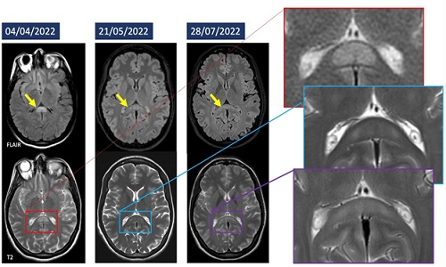 Figure 1: Balance and subjective visual disturbances of the patient.
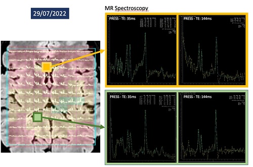 Figure 2: MRI showed a reduction in lesion size, although MR spectroscopy showed a decrease in metabolites levels in the splenium.
Acknowledgement
Partially funded by ricerca corrente linea 2, italian ministers of Health.
References
- Lewis R, Ruiz A, Monteith T (2020) Reversible Lesion of the Corpus Callosum in a Patient With Migraine With Aura: A Case Study Headache. 60: 791-792.
- Tetsuka S (2019) Reversible lesion in the splenium of the corpus callosum. Brain Behav. 9: e01440.
Citation: Fusco FR, Bizzaglia M, Luccichenti G (2023) Cytotoxic Lesion of the Corpus Callosum: A Case of Metabolic Disease in a New Mother. J Clin Stud Med Case Rep 10:208
Copyright: © 2023 Francesca R Fusco, et al. This is an open-access article distributed under the terms of the Creative Commons Attribution License, which permits unrestricted use, distribution, and reproduction in any medium, provided the original author and source are credited.