DNA Interaction Study of Some Symmetrical 1,2-Phenylenediamine Schiff’s Base Derivatives as New Potential DNA Intercalators using Ethidium Bromide Competition Fluorescent Assay
*Corresponding Author(s):
Gbaj AMAssociate Professor Of Genetics And Biochemistry, Department Of Medicinal Chemistry, Faculty Of Pharmacy, University Of Tripoli, Libyan Arab Jamahiriya
Tel:+218 913556785,
Email:abdulgbaj4@hotmail.com
Abstract
Keywords
INTRODUCTION
Intercalators are ligands interacting with the DNA double helix in a reversible manner. Many of them are currently used as powerful drugs for the treatment of breast and ovarian cancers, acute leukemia, and many others are still in clinical trials. Intercalating agents have several universal structural features; the most important is a planar polyaromatic system that inserts between DNA base-pairs with a marked preference for the purine-3'-pyrimidine-5' sequence [10-12]. Ethidium bromide is frequently utilized as a fluorescent tag (nucleic acid stain) in molecular biology for many procedures such as agarose gel electrophoresis. It intercalates with double-stranded DNA and leads to the deformation of the DNA which could affect biological processes, such as DNA transcription and replication [13,14]. Schiff’s bases (imines) are compounds that contain azomethine groups [-HC=N-] in their structure. They are formed by condensation of a dynamic carbonyl compound with a primary amine [15,16]. Schiff’s bases show a large range of biological activities including anti-viral, anti-bacterial, anti-inflammatory, anti-proliferative, antifungal, anti-malarial and antipyretic pharmacological activities [17-21]. We have recently reported on using microwave assisted synthesis and antimicrobial evaluation of symmetrical 1,2-Phenylenediamine Schiff’s base derivatives finding that some of the synthesized compounds showed antibacterial and antifungal activity [22]. Now our aim is to explore how different structural features of 1,2-Phenylenediamine Schiff’s base derivatives can affect the DNA binding capability using molecular modeling and key selection vector studies which may serve as a basis for understanding the molecular mechanism of action of the 1,2-Phenylenediamine Schiff’s base derivatives and can help to design of new chemotherapeutic molecules.
MATERIAL AND METHODS
Chemistry
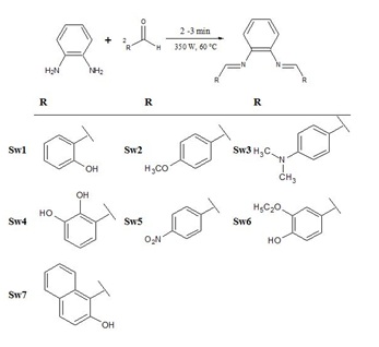
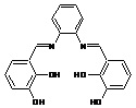
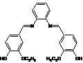
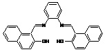
Schiff’s bases were prepared according to our previously reported procedure by reacting of one mole of phenylenediamine and two moles of substituted aromatic aldehydes [22]. All reactants were mixed together and small amount of ethanol (5-6 ml) was added. This mixture was subjected to microwave irradiation at 350 watt for 2-3 minutes at a maximum of 60°C. After the reaction was completed (2-3 minutes), the mixture was left for cooling and the solid product (crude) was gathered by filtration and washed several times (5-6 washes) with ethanol and then vacuum dried. The gained product was recrystallized in ethanol and after drying that gave the clean pure product (Scheme 1). The crystalline product obtained was characterized as described previously by FTIR and 1H NMR [22].
To study how competently our synthesized compounds interact with G-DNA, the compounds were investigated for their DNA binding ability using fluorescence emission spectroscopy. All experiments were conducted in Tris buffer (0.01M Tris, 0.1M NaCl, at pH 7.4), glass-distilled deionized water and analytical grade reagents were used throughout experiments. The pH values of all solutions were measured with a calibrated Jenway pH-meter model 3510 (Staffordshire, UK). All buffer solutions were filtered through Millipore filters (Millipore, UK) of 0.45 mm pore diameter.
Absorbance spectra
Absorbance spectra were measured on a Jenway UV-visible spectrophotometer, model 6505 (London, UK) using quartz cells of 1.00 cm path length. The UV-Vis absorbance spectra were recorded in the 200-500nm range and a spectral bandwidth of 3.0 nm. For the final spectrum of each solution analyzed baseline subtraction of the buffer solution was performed. Genomic DNA was used in a concentration of 75μg/ml. DNA was extracted from peripheral lymphocytes of anti-coagulated blood (EDTA) samples by Proteinase K digestion and phenol/chloroform extraction [23]. The purity was determined by measuring the absorbance at 260/280nm indicating that the sample is free from protein contamination [23]. The concentration was assayed spectrophotometrically using 6600M-1cm-1 as a molar extinction coefficient at 260nm.
Fluorescence spectra and DNA-binding studies
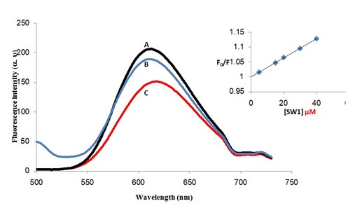
Fluorescence emission and excitation spectra were measured using Jasco FP-6200 spectrofluorometer (Tokyo, Japan) using fluorescence 4-sided quartz cuvettes of 1.00cm path length. The automatic shutter-on function was used to minimize photo bleaching of the sample. The selected excitation wavelength for ethidium bromide was 480 nm. The emission spectrum was corrected for background fluorescence of the buffer. The Ethidium Bromide (EB) fluorescence displacement experiment was performed by sequential addition of aliquots of 1790μl Tris buffer, 10μl EB (final concentration of 72μM), 100μl G-DNA from stock solutions (1.5mg/ml) and finally 10μl of compounds (SW1 to SW7, final concentration of 30μM). Emission spectra were recorded for each system using excitation wavelengths of maximum fluorescence intensity determined for the systems to be 480nm using a slit width of 5nm to examine alterations in emission spectra resulting from the complex construction of both systems. On construction of the full systems, the system was allowed to equilibrate for 30 minutes at room a temperature and emission spectra (500-730nm) were recorded to monitor changes in EB intensity.
Prediction of ADMET properties and drug-likeliness
To verify drug-likeliness, canonical SMILES format of the synthesized molecules SW compounds were used in swiss ADME (http://www.swissadme.ch/index.php) [24]. The main bioavailability parameters were molecular weight (g/mol), lipophilicity (XlogP3), solubility (log S), polarity (Topological Polar Surface Area-TPSA in Å2), saturation (fraction of carbon atoms in Sp3 hybridization-Csp3) and flexibility (No of rotatable bonds). Absorption parameters such as Human Intestinal Absorption, Blood Brain Barrier, P-glycoprotein interaction (substrate or inhibitor) and metabolism (inhibitor or substrate) of the bio-active molecules with different cytochrome P450 enzymes were also estimated using admetSAR, which is based on QSAR data for prediction of Absorption, Distribution, Metabolism, Excretion and Toxicity (ADMET) [25,26]. Drug-like molecules considering Ghose, Veber, Egan, Muegge and Lipinski rules and good ADMET properties were chosen as ligands in a consequent molecular docking procedure.
Molecular docking
The starting geometry of the synthesized SW compounds (SW1 to SW7) was constructed using chem3D Ultra (version 8.0, Cambridge soft Com., USA). The optimized geometry of SW compound with the lowest energy was used for molecular docking. Crystal structures of the DNA fragment with the B-form 1bna, 102d, 1k2j and 102d were downloaded from the Protein Data Bank (https://www.rcsb.org/structure/102D). Molecular dockings of SW compounds with B-DNAs were accomplished by Auto Dock 4.2 software from The Scripps Research Institute (TSRI) (http://autodock.scripps.edu/). Firstly, polar hydrogen atoms were added into B-DNA molecules. Then, partial atomic charges of the B-DNA and SW molecules were calculated using Kollman methods [27]. In the process of molecular docking, the grid maps of dimensions (62Å × 62Å × 62Å) with a grid-point spacing of 0.376Å and the grid boxes are centered. The number of genetic algorithm runs and the number of evaluations were set to 100. All other parameters were default settings. Cluster analysis was performed on the results of docking by using a Root Mean Square (RMS) tolerance of 2.0Å, which was dependent on the binding free energy. Lastly, the dominating configuration of the binding complex of SW compounds and B-DNA fragments with minimum energy of binding were determined which relied strongly on the information of 3D structures of the DNA binding site and ultimately generated a series of DNA binding complex.
RESULTS AND DISCUSSION
Synthesis
ADMET properties
|
Compounds (M Wt) g/mol |
Chemical structure |
Log P* |
Hydrogen bonds** |
KSV (M-1) |
R2 (5 points) |
|
|
Calculated |
donors |
acceptors |
||||
|
Taxol (853.92) |
|
2.5 |
4 |
14 |
5.90 × 10³ |
0.999 |
|
SW1 (316.35) |
|
4.99 |
2 |
4 |
3.17 × 10³ |
0.968 |
|
SW2 (344.4) |
|
5.52 |
0 |
4 |
1.10 × 10³ |
0.995 |
|
SW3 (370.49) |
|
6.34 |
0 |
2 |
1.20 × 10³ |
0.987 |
|
SW4 (348.35) |
 |
4.21 |
4 |
6 |
6.10 × 10³ |
0.996 |
|
SW5 (374.34) |
|
5.84 |
0 |
6 |
6.02 × 10³ |
0.988 |
|
SW6 (404.45) |
 |
5.41 |
2 |
6 |
4.7 × 10³ |
0.996 |
|
SW7 (416.47) |
 |
6.98 |
2 |
4 |
7.31 × 10³ |
0.998 |
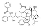
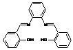
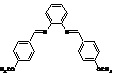
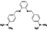
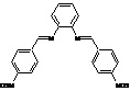
Table 1: Some chemical, physical properties and the key selection vector (KSV) values of compounds (SW1-SW7).
Note: *Log P values were calculated using ChemDraw Ultra 8.0 software. **Numbers and type of hydrogen bonds involved in the interaction between SW compounds and DNA were determined using swiss ADME website [24].
Molecular docking analysis
|
Compounds (Ligands)
|
DNA PBD ID(1bna) |
DNA PBD ID (102d) |
DNA PBD ID(1k2j) |
DNA PBD ID (102d) |
|
ΔGa |
ΔGa |
ΔGa |
ΔGa |
|
|
Sw1 |
-7.8 |
-5.73 |
-5.03 |
-5.61 |
|
Sw2 |
-6.4 |
-6.67 |
-7.57 |
-5.2 |
|
Sw3 |
-6.7 |
-5.03 |
-5.23 |
-6.19 |
|
Sw4 |
-8.0 |
-5.73 |
-6.0 |
-5.51 |
|
Sw5 |
-7.4 |
-3.29 |
-6.78 |
-3.21 |
|
Sw6 |
-6.8 |
-4.58 |
-4.92 |
-5.27 |
|
Sw7 |
-7.9 |
-6.02 |
-5.39 |
-6.94 |
|
Taxol |
-1.16 |
-3.24 |
-12.38 |
-3.65 |
The molecular modeling results showed that there are van der Waals, hydrogen bonds and electrostatic interactions between ethidium bromide, SW compounds and DNA. The contribution of van der Waals and hydrogen bond interaction is much greater than that of the electrostatic interaction because the sum of van der Waals energy, hydrogen bonding energy and desolvation free energy is larger than the electrostatic energy, which is consistent with the experimental results shown in Table 1. As indicated in the literature, the binding mode of ethidium bromide on DNA is intercalating binding [28]. The fluorescence emission intensity of ethidium bromide decreased on addition of SW compounds suggesting that there is competitive binding between ethidium bromide and SW compounds on G-DNA. Therefore, it can be concluded that the binding mode of ethidium bromide on G-DNA is intercalating binding which is consistent with our modeling and fluorescence results.
DNA Binding Properties
Ethidium bromide (EB) Competition Assay
CONCLUSION
AUTHOR CONTRIBUTION
ACKNOWLEDGEMENT
CONFLICT OF INTEREST
ETHICAL APPROVAL
REFERENCES
- Hartley JA, Hochhauser D (2012) Small molecule drugs-optimizing DNA damaging agent-based therapeutics. Curr Opin Pharmacol 12: 398-402.
- Srinivasan A, Gold B (2012) Small-molecule inhibitors of DNA damage-repair pathways: An approach to overcome tumor resistance to alkylating anticancer drugs. Future Med Chem 4: 1093-1111.
- Hamilton PL, Arya DP (2012) Natural product DNA major groove binders. Nat Prod Rep 29: 134-143.
- Denny WA (2001) DNA minor groove alkylating agents. Curr Med Chem 8: 533-544.
- Lucas MF, Cabeza de Vaca I, Takahashi R, Rubio Martinez J, Guallar V (2014) Atomic level rendering of DNA-drug encounter. Biophys J 106: 421-429.
- Xi H, Davis E, Ranjan N, Xue L, Hyde Volpe D, et al., (2011) Thermodynamics of nucleic acid "shape readout" by an amino sugar. Biochemistry 50: 9088-9113.
- Fairbanks SD, Robertson CC, Keene FR, Thomas JA, Williamson MP (2019) Structural Investigation into the threading intercalation of a Chiral Dinuclear Ruthenium(II) Polypyridyl Complex through a B-DNA Oligonucleotide. J Am Chem Soc 141: 4644-4652.
- Bera D, Verdonck L, Glassner M, Madder A, Hoogenboom R (2019) Thermoresponsive DNA by Intercalation of dsDNA with Oligoethylene-Glycol-Functionalized small-molecule intercalators. Macromol Rapid Commun 40: e1800900.
- Zanoza SO, Klimenko KO, Maltzev GV, Bykova TI, Levandovskiy IA, et al., (2019) Aminoalkoxyfluorenones and aminoalkoxybiphenyls: DNA binding modes. Bioorg Chem 86: 52-60.
- Schonn I, Hennesen J, Dartsch DC (2010) Cellular responses to etoposide: Cell death despite cell cycle arrest and repair of DNA damage. Apoptosis 15: 162-172.
- Ross WE, Bradley MO (1981) DNA double-stranded breaks in mammalian cells after exposure to intercalating agents. Biochim Biophys Acta 654: 129-134.
- Tewey KM, Rowe TC, Yang L, Halligan BD, Liu LF (1984) Adriamycin-induced DNA damage mediated by mammalian DNA topoisomerase II. Science 226: 466-468.
- Gao Y, Li J, Huang G, Yan L, Dong Z (2015) Spectroscopic studies on the interaction between anthragallol and DNA using of ethidium bromide as a fluorescence probe. Spectrochim Acta A Mol Biomol Spectrosc 141: 239-243.
- Zhang G, Hu X, Fu P (2012) Spectroscopic studies on the interaction between carbaryl and calf thymus DNA with the use of ethidium bromide as a fluorescence probe. J Photochem Photobiol B 108: 53-61.
- Thies H, Schonenberger H, Bauer Kh (1958) [Reactions of Schiff's bases. V. Transformation of benzylidene-arylamines with mixtures of magnesium and magnesium iodide]. Arch Pharm Ber Dtsch Pharm Ges 291/63: 620-627.
- Puchtler H, Meloan SN, Brewton BR (1975) On the history of basic fuchsin and aldehyde-Schiff reactions from 1862 to 1935. Histochemistry 41: 185-194.
- Piscopo E, Diurno MV, Cirino G, Antonucci M, Cataldi MT, et al., (1984) [Biological activity of 4-hydroxy-5-formylbenzoic acid derivatives. II. Esters and Schiff bases with antimicrobial activity]. Boll Soc Ital Biol Sper 60: 501-507.
- Lubec G, Leban J, Peyroux J, Sternberg M, Pollak A, et al., (1982) Reduced collagenolytic activity of rat kidneys with steptozotocin diabetes. Nephron 30: 357-360.
- Rahman AH, Ismail EM (1976) Synthesis of Schiff bases of benzofuran with potential biological activity. Arzneimittelforschung 26: 756-759.
- Lamie PF, Ali WAM, Bazgier V, Rarova L (2016) Novel N-substituted indole Schiff bases as dual inhibitors of cyclooxygenase-2 and 5-lipoxygenase enzymes: Synthesis, biological activities in vitro and docking study. Eur J Med Chem 123: 803-813.
- Meng XY, Liu JQ, Zhang XP, Chen XH, Yu AZ, et al. (1994) [Synthesis and biological activities of 2,4-diamino-5-fluoro-6-substituted benzylamino quinazolines]. Yao Xue Xue Bao 29: 261-267.
- Mohamed SS, Sadawi IAA, Gbaj MA, Alsabri SG, Elmaki NM, et al., (2018) Microwave Assisted Synthesis and Antimicrobial Evaluation of Symmetrical 1,2-Phenylenediamine Schiff's Base derivatives. Pharm Pharmacol Int J 6: 344-348.
- Johns MB Jr., Paulus Thomas JE (1989) Purification of human genomic DNA from whole blood using sodium perchlorate in place of phenol. Anal Biochem 180: 276-278.
- Daina A, Michielin O, Zoete V (2017) SwissADME: A free web tool to evaluate pharmacokinetics, drug-likeness and medicinal chemistry friendliness of small molecules. Sci Rep 7: 42717.
- Guan L, Yang H, Cai Y, Sun L, Di P, et al. (2019) ADMET-score - a comprehensive scoring function for evaluation of chemical drug-likeness. Medchemcomm 10: 148-157.
- Yang H, Lou C, Sun L, Li J, Cai Y, et al., (2019) admetSAR 2.0: Web-service for prediction and optimization of chemical ADMET properties. Bioinformatics 35: 1067-1069.
- Tiwari R, Mahasenan K, Pavlovicz R, Li C, Tjarks W (2009) Carborane clusters in computational drug design: A comparative docking evaluation using AutoDock, FlexX, Glide, and Surflex. J Chem Inf Model 49: 1581-1589.
- Krishna AG, Kumar DV, Khan BM, Rawal SK, Ganesh KN (1998) Taxol-DNA interactions: Fluorescence and CD studies of DNA groove binding properties of taxol. Biochim Biophys Acta 1381: 104-112.
- Kuruvilla E, Joseph J, Ramaiah D (2005) Novel bifunctional acridine-acridinium conjugates: Synthesis and study of their chromophore-selective electron-transfer and DNA-binding properties. J Phys Chem B 109: 21997-22002.
- Tsuboi M, Benevides JM, Thomas GJ (2007) The Complex of ethidium bromide with genomic DNA: Structure analysis by polarized raman spectroscopy. Biophys J 92: 928-934.
- Zhang QQ, Zhang F, Wang WG, Wang XL (2006) Synthesis, crystal structure and DNA binding studies of a binuclear copper(II) complex with phenanthroline. J Inorg Biochem 100: 1344-1352.
- Jiang M, Li YT, Wu ZY, Liu ZQ, Yan CW (2009) Synthesis, crystal structure, cytotoxic activities and DNA-binding properties of new binuclear copper(II) complexes bridged by N, N'-bis(N-hydroxyethylaminoethyl) oxamide. J Inorg Biochem 103: 833-844.
- Prunkl C, Pichlmaier M, Winter R, Kharlanov V, Rettig W, et al., (2010) Optical, redox, and DNA-binding properties of phenanthridinium chromophores: Elucidating the role of the phenyl substituent for fluorescence enhancement of ethidium in the presence of DNA. Chemistry 16: 3392-3402.
- Garbett NC, Hammond NB, Graves DE (2004) Influence of the amino substituents in the interaction of ethidium bromide with DNA. Biophys J 87: 3974-3981.
- Li H, Yu YY, Hu X, Cao SW (2008) [Research on the interactions between genistein and its glucosides with DNA]. Guang Pu Xue Yu Guang Pu Fen Xi 28: 1905-1909.
- Yu YY, Li H, Hu X, Cao SW (2008) [Research on the interaction of Cr(III) complex of genistein with DNA]. Guang Pu Xue Yu Guang Pu Fen Xi 28: 1587-1591.
- Dezhampanah H, Fyzolahjani S (2013) Study on interaction of cationic porphyrazine with synthetic polynucleotides. Anal Cell Pathol (Amst) 36: 125-132.
Citation: Mohamed SS, Sadawe IA, Meiqal NH, Alshoushan AA, Aboud AS, et al. (2019) DNA Interaction Study of Some Symmetrical 1,2-Phenylenediamine Schiff’s Base Derivatives as New Potential DNA Intercalators using Ethidium Bromide Competition Fluorescent Assay. J Mod Chem Sci 3: 005.
Copyright: © 2019 Mohamed SS, et al. This is an open-access article distributed under the terms of the Creative Commons Attribution License, which permits unrestricted use, distribution, and reproduction in any medium, provided the original author and source are credited.