Early Multidisciplinary Approach for the Management of Dilacerated Permanent Maxillary Incisor: A Case Report
*Corresponding Author(s):
Wang JingDepartment Of Stomatology, The First Medical Center Of PLA General Hospital, Beijing, China
Email:wangjplagh@sina.com
Abstract
Management of dilaceration teeth poses a unique challenge to the clinician due to its position within the esthetic zone. This case report describes the management of impacted maxillary central incisor with severe root dilacerations. Surgical exploration was done and orthodontic treatment was planned. The dilacerated incisor was alighed, optimal treatment outcome with esthetic and functional was well done.
Keywords
Dilaceration; Impacted; Orthodontic treatment; Surgical exploration
Background
Tooth dilaceration is a developmental disturbance that is associated with a change in orientation of the root often in the labiolingualor mesiodistal direction. John Tomes in 1848 was the first person to report a central incisor with angulated root and coined it as ”Dilaceration” [1]. He considered it as aforcible separation of developed dentin from the developing dentin. Prevalence was reported to be 4.6%. The etiological factors for the impacted incisors can be because of trauma, mechanical obstruction by supernumerary teeth, developmental disturbances.
A 8-year-old boy reported to the Department of Orthodontic Dentistry with a complaint of missing upper front tooth. History revealed that the patient had an injury to the front tooth region 4 years ago, due to a fall. Intraoral examination revealed a missing right central incisor without space loss (Figure 1A). CBCT revealed an impacted malformed central incisor which necessitated further investigations (Figure 1B and 1C). To assess the detailed three-dimensional positioning of the root in orthogonal and oblique planes, cone-beam computed tomography was advised, which revealed dilaceration root with more than 90° angulation and crown directed toward the ANS (Figure 1B and 1 C).
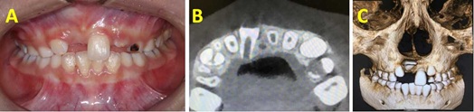 Figure 1 (A to C): A) Intraoral photograph. B and C) CBCT showing impacted dilacerated central incisor.
The apical foramen of impacted malformed central incisor was not closed, guided eruption combined with surgical exploration was possible. Lingual button was bonded on lingual side of the incisor during surgical flap operation. The patient was recalled after 1 week and orthodontic traction was taken. Removable appliances was used for 3 months. Interactive traction was used for 2 months. Fixed orthodontic treatment was planned to align the upper anterior teeth for 3 months (Figure 2). Lingual retainer was bonded from letaral incisor to letaral incisor (Figure 3).
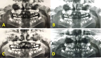 Figure 2 (A to D): Panoramic radiography showing the movement of impacted dilacerated central incisor during orthodontic treatment.
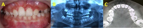
Figure 3 (A to C): 3A) Intraoral photograph after treatment (B to C) Panoramic radiography and CBCT showing the correct position of incisor.
Discussion
Impaction of incisors is not a frequent event in dental practice. Most orthodontists believe that the primary etiological factor for dilacerated incisor is trauma [2,3]. However, other factors do exist and the exact cause may not be fully understood.
It is an arduous clinical task for the rehabilitation of dilacerated impacted central incisor. Factors should be considered for successful treatment include: Crown root alignment of the impacted tooth; Stage of root development; Deviated angle of dilaceration; Space present in arch for aligning the impacted tooth.
Impacted central and lateral incisors can be managed in three divided phases. Pre-surgical orthodontic measures first create sufficient space for the unerupted tooth. Surgical uncovering of the impacted too this followed by gingivectomy ordisplacedflap [4]. Orthodontic traction and alignment in the post-surgical orthodontic phase complete the treatment.
For a better prognosis of orthodontic traction, the position of the dilacerated root and its developmental stage plays a pivotal role. The positioning of the incisors was obtained by creating the eruptive space and aligning the un-erupted teeth with orthodontic treatment or after removing any obstacle or after their exposure with different surgical methods [5].
In the present case report to appreciate these features, CBCT revealed that the dilaceration was more than 90° toward the crown, and the crown was directed toward the ANS. Previous studies stated that orthodontic traction of severely dilacerated teeth may result in ankylosis [6], periodontal attachment loss, root resorption, and exposure. However, the present analysis shows that the root of the impacted incisor could achieve better development if treated earlier [3]. We believed that light force traction is helpful for root shaping of apical unclosed teeth because no prosthetic solutions is better than the tooth itself, as the volume of alveolar bone is preserved [7].
A removable appliance was preferred firstly as it is easy to wear and maintain the oral hygiene. After crown eruption, lingual buttons was changed to labial surface and bonded to establish a couple of forces with lower anterior teeth. In the occlusal adjustment stage, a fixed appliance was preferred as it minimizes patient discomfort, reduces the need for patient compliance with increased control of tooth movements in all three directions of space.
Conclusion
In this case, surgical and orthodontic treatments were sequentially planned for optimal treatment outcome with esthetic and functional well-being.
Clinical Significance
Accurate assessment of the early symptoms of an affected tooth is crucial for proper diagnosis and for establishing a comprehensive treatment plan which minimizes future adverse effects. Guided eruption combined with surgical exploration was possible for anterior dilacerated tooth with unclosed apical foramen.
References
- Nallanchakrava S, Mettu S, Reddy NG, Jangam K (2020) Multidisciplinary approach for the management of dilacerated permanent maxillary incisor: A case report. Int J Clin Pediatr Dent 13: 725-728.
- Goswami M, Bhardwaj S (2022) Assessment of traumatic dental injuries among institutionalized orphan children: A cross-sectional study. Int J Clin Pediatr Dent 15: 124-127.
- Žaroviene A, Grinkeviciene D, Trakiniene G, Smailiene D (2021) Post-treatment status of impacted maxillary central incisors following surgical-orthodontic treatment: A systematic review. Medicina 57: 783.
- Ahammed H, Seema T, Deepak C, Ashish J (2021) Surgical management of impacted supernumerary tooth: A case series. Int JClin Pediatr Dent 14: 726-729.
- Bhoila R, Bramtab M, Bhoi R (2020) Bull horn injury causing traumatic tooth intrusion - ultrasound and CT imaging. Afr J Emerg Med 10: 99-102.
- Migliario M, Lucchina AG, Rocchetti V, Renò F (2019) Laser surgical approach to impacted maxillaryincisors: Case series and brief review. Eur Rev Med Pharmacol Sci 23: 9691-9696.
- Mahardawi B, Kumar KC, Arunakul K, Chaiyasamut T, Wongsiricha N (2020) Judgement in artificial eruption of embedded teeth froman oral surgery perspective: Review article. J Korean Assoc Oral Maxillofac Surg 46: 121-18.
Citation: Jing W, Naand H, Chuan C (2023) Early Multidisciplinary Approach for the Management of Dilacerated Permanent Maxillary Incisor: A Case Report. Int J Case Rep Ther Stud 4: 019.
Copyright: © 2023 Wang Jing, et al. This is an open-access article distributed under the terms of the Creative Commons Attribution License, which permits unrestricted use, distribution, and reproduction in any medium, provided the original author and source are credited.