Esophageal Plexiform Fibromyxoma: An Unusual Localization
*Corresponding Author(s):
Lorenzo Del NeroSC Gastroenterologia ASL 2 Savonese, Ospedale Santa Corona, Pietra L (SV), Italy
Email:l.delnero@asl2.liguria.it
Introduction
Plexiform Fibromyxoma (PF) is an infrequent mesenchymal tumor of the Gastrointestinal Tract (GI). The reported incidence of PF relative to GI Stromal Tumor (GIST) is estimated 1.7 % over a time of 20 year [1].
PF is mostly found in the gastric antrum. However, it has been described in other GI tract segments such as duodenum, jejunum, gallbladder and mediastinum [2]. Esophageal presentation is extremely rare.
A case report of an esophageal PF is described in a 55-year-old woman.
Case Report
A 55-year-old woman with a two-year history of mild dysphagia was referred to our Endoscopy Center for evaluation of a proximal esophageal polyp found in a previous esophagogastroduodenoscopy.
A CT scan of the neck revealed thickening of the cervical esophagus in the absence of other pathological finding (figure 1).
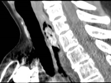 Figure 1: CT image of the esophageal polyp
An endoscopic re-evaluation confirmed a 30 mm sessile polyp with high vascularity. Endoscopic Submucosal Dissection (ESD) of the lesion was performed without immediate or late adverse events.
Macroscopic examination revealed a 3.1 x 2.6 cm lesion with a bozzellated and partly hemorrhagic surface.
The pathological features were a multinodular proliferative lesion, composed of bland, uniform spindle-ovoid cells with no mitotic activity, in richly vascularized interstizial mixoid matrix with focal chronic inflammatory chances. The proliferative index (MIB1/Ki67) was 1%. Immunohistochemistry: neoplastic cells express smooth muscle actin. S100, H caldesmon, CD117, DOG1, CD34, SOX10, CKAE1-AE3 GFAP, MUC4, MDM2 were negative (Figures 2,3,4). Strikingly the histo-cytopathological analysis led to diagnosis of plexiform fibromyxoma.
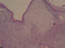 Figure 2: Hematoxylin eosin staining
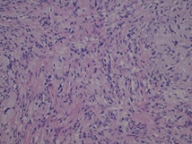 Figure 3: Hematoxylin eosin staining
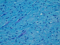 Figure 4: Alcian PAS
Discussion
Due to its rarity, not many cases of PF have been described in literature. In particular, esophageal presentation is extremely rare.
Pathogenesis and molecular alterations of PF are largely unknown and its incidence seems to be increasing in the last years [3].
PF is equally described in males and females and the median age at presentation is 40 years to 50 years, although pediatric cases have been described as well [4,5].
In the majority of patients, the clinical presentation is not specific: abdominal pain, early fullness, vomiting and anemia are sometimes present.
Due to the submucosal localization of the tumor, EUS + FNA are considered the diagnostic gold standards, even if primary resection is usually the first choice [6].
The pathologist's approach to gastroesophageal mesenchymal tumors has been deeply reviewed in the last years thanks to increasingly detailed genetic subclassification. Specific treatments optimized for particular genetic subtypes are now available [7].
PF are usually considered benign conditions, even if vascular, lymphatic and mucosal invasion have been described. Neoplasia with similar histologic features (GIST, smooth muscle tumors) have instead a malignant potential [1]. For this reason, an accurate differential diagnosis is essential [6].
References
- Arslan ME, Li H, Jennings TA, Lee EC, Nigam A, et al. (2020) Frequency of Plexiform Fibromyxoma relative to gastrointestinal stromal tumor: A single center study. Ann Diagn Pathol 48: 151568.
- Arslan ME, Li H, Fu Z, Jennings TA, Lee H (2021) Plexiform fibromyxoma: Review of rare mesenchymal gastric neoplasm and its differential diagnosis. World J Gastrointest Oncol 13: 409-423.
- Takahashi Y, Suzuki M, Fukusato T (2010) Plexiform angiomyxoid myofibroblastic tumor of the stomach. World J Gastroenterol 16: 2835-2840.
- Su HA, Yen HH, Chen CJ (2019) An Update on Clinicopathological and Molecular Features of Plexiform Fibromyxoma. Can J Gastroenterol Hepatol 2019: 3960920.
- Duckworth LV, Gonzalez RS, Martelli M, Liu C, Coffin CM, et al. (2014) Plexiform fibromyxoma: report of two pediatric cases and review of the literature. Pediatr Dev Pathol 17: 21-27.
- Álvarez MC, Martínez JMO, Martínez ET, Carrera MST, Díaz MJF, et al. (2021) Gastric plexiform fibromyxoma, an uncommon mesenchymal tumor. Rev Esp Enferm Dig 113: 183-185.
- Papke DJ, Hornick JL (2021) Recent developments in gastroesophageal mesenchymal tumours. Histopathology 78: 171-186.
Citation: Nero LD, Ziola S, Dellachà A, Quilici P, Ceglie AD, et al. (2023) Esophageal Plexiform Fibromyxoma: An Unusual Localization. J Gastroenterol Hepatology Res 7: 046
Copyright: © 2023 Lorenzo Del Nero, et al. This is an open-access article distributed under the terms of the Creative Commons Attribution License, which permits unrestricted use, distribution, and reproduction in any medium, provided the original author and source are credited.