Introducing a New Term: Metastasoptysis- A Case Report
*Corresponding Author(s):
Sercan AydinDepartment Of Thoracic Surgery, School Of Medicine, Ege University, Izmir, Turkey
Tel:+90 5069332545,
Email:drsrcn@gmail.com
Abstract
Pulmonary metastasis is not uncommon in the follow-up of patients with primary malignancy, but the expectoration of metastatic tumor is extremely rare. Metastases from renal and breast malignities may rarely lead to tumor expectoration with less common colorectal carcinomas. In this case report, expectoration of a metastatic tumor in a patient who had diagnosed with colon adenocarcinoma and pulmonary metastasis was presented. We named this quite rare condition as “metastasoptysis”.
Keywords
Expectoration; Metastasoptysis; Pulmonary metastasis
INTRODUCTION
Pulmonary parenchyma is among the target organs of most metastatic diseases. Although most of the patients are asymptomatic; chest pain, dyspnea, hemoptysis, and cough may be encountered [1]. Very rarely, metastases can be expectorated after severe cough attacks [2].
CASE REPORT
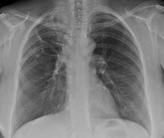
A 51-year-old female who had undergone colon resection and pulmonary metastasectomy16 months ago due to colon adenocarcinoma and pulmonary metastasisadmitted to our outpatient clinic for annual control. Radiologic examination revealed usual postoperative changes, such as focal opacity and loss of aeration in the upper zone of right lung secondary to the surgery (Figure1).
 Figure 1: The chest posteroanterior radiograph of the patient at admission.
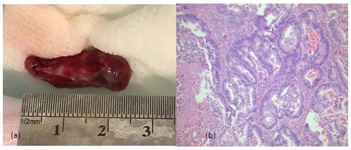
Laboratory findings were in the normal range. On the first day of admittance, she had a severe cough attack. The patient who did not complain of nausea expectorated a reddish mucous solid material having the dimensions of 2.5x1x0.6 cm (Figure 2a). No bleeding or mass was found in the mouth and throat examination after the coughing attack. The patient stated that there was no previous lesion in her mouth.Histopathological examination of the expectorated material showed irregular branching, anastomosing and cribriform glands with columnar epithelium. The columnar cells had pseudostratified vesicular nuclei. The glandular lumen contained nuclear debris and necrotic cells (i.e.dirty necrosis) and reported as adenocarcinoma with the same histopathological structure as the pulmonary metastasectomy material examined before (Figure 2b). The patient was referred to the Medical Oncology Clinic for chemotherapy. After metastasoptysis, 2 cycles of chemotherapy was applied to the patient who was decided to be followed up in the postoperative period. But the patient died approximately 6 months after the second course of chemotherapy.
 Figures 2(a,b): a. Expectorated material, b. Adenocarcinoma showing complex glandular structures and luminal necrotic debris (dirty necrosis). (H&E stain x 100).
DISCUSSION
Although the most common tumor expectoration cases are due to metastasis in the literature, cases in which primary lung carcinoma masses are expectorated have also been reported. Squamous cell carcinomas and bronchogenic carcinomas are more common than other primary lung carcinoma histopathological types [3]. Re-development of lung metastasis in patients after pulmonary metastasectomy is a common phenomenon [1,4]. Rarely, the tumor tissue can be expectorated by the patient. Tumors removed by expectoration have different histopathologies in the literature. No specified data on the fragility or size of the tumor were found in publications. Their common point is that it is inside the airways or protrudes into the bronchus by compressing from the outside [3,5]. In the English literature, there were two reported cases of malignant tumor expectoration: one was 18 months after the upper left lobectomy due to primary lung adenocarcinoma, whereas the other was in a patient diagnosed with colon adenocarcinoma who developed a lung metastasis during the follow-up [3,6]. Our patient expectorated metastasis 16 months after the operation, as similar to the first reported patient. The latter one had not undergone lung surgery unlike the present case. We believe that this is the third case in the literature. In order to bring a new term into the medical literature, we want to define metastatic tumor tissue expectoration as "metastasoptysis". We can also say that in patients having malignant disease, any expectorated material should be examined histopathologically.
CONFLICT OF INTEREST
The authors declared no conflicts of interest with respect to the authorship and/or publication of this article.
FUNDING
The authors received no financial support for the research and/or authorship of this article. The procedures followed were in accordance with the ethical standards of the responsible committee on human experimentation (institutional and national) and with the Declaration of Helsinki.
REFERENCES
- Allen MS, Putnam JB. Secondary Tumors of the Lung. In: Shields TW, LoCicero III J, Reed CE and Feins RH (eds). General Thoracic Surgery, Seventh ed. Lippincott Williams &Wilkins, Philadelphia, 2009: 1619-1646.
- Heitmiller RF, Marasco WJ, Hruban RH (1993) Endobronchial metastasis. J Thorac Cardiovasc Surg 106: 537-542.
- Ochi N, Yasugi M, Yamane H, Tabayashi T, Kawanaka N, et al. (2012) Spontaneous expectoration of tumor tissue in a patient with adenocarcinoma of the lung. Respir Care 57: 1521-1523.
- Lee W, Yun SH, Chun HK, et al. (2007) Pulmonary resection for metastases from colorectal cancer: prognostic factors and survival. Int J Colorectal Dis 22: 699-704.
- Kern WH (1976) Expectoration of Bronchogenic Tumor Tissue. JAMA 236: 2604.
- Ghetie C, Davies M, Cornfeld D, Suh N, Wasif Saif M (2008) Expectoration of a lung metastasis in a patient with colorectal carcinoma RSS. Clin Colorectal Cancer 7: 283-286.
Citation: Aydin S, Cakan A, Cagirici U, Ozyigit Buyuktalanci D (2020) Introducing a New Term: Metastasoptysis- A Case Report. Int J Case Rep Ther Stud 2: 12
Copyright: © 2020 Sercan Aydin, et al. This is an open-access article distributed under the terms of the Creative Commons Attribution License, which permits unrestricted use, distribution, and reproduction in any medium, provided the original author and source are credited.

