Journal of Otolaryngology Head & Neck Surgery Category: Clinical
Type: Case Report
Melanotrichoblastoma of Nose: A Case Report
*Corresponding Author(s):
Bouatay REnt Department, Fattouma Bourguiba Hospital At Monastir, University Of Monastir, Tunisia
Tel:+216 24066013,
Email:rbouattay@yahoo.fr
Received Date: Jun 01, 2019
Accepted Date: Jun 21, 2019
Published Date: Jun 28, 2019
Abstract
Melanotrichoblastoma represents a rare variant of trichoblastoma in which neo plastic epithelium is colonized by dendritic melanocytic cells. We report in this paper a rare case of melanotrichoblastoma located in the nose, diagnosed in a 67-year-old female.
Keywords
Epithelial cells; Melanocytes; Melanotrichoblastoma; Trichoblastoma
INTRODUCTION
Trichoblastoma are benign skin adnexal tumors, under the category of trichogenic tumors. It shows proliferation of epithelial and mesenchymal cells which recapitulate the hair follicle development [1]. Melanotrichoblastoma is a rare variant of pigmented trichoblastoma. Only few cases have been reported in literature [2]. We report a rare case of melanotrichoblastoma of nose and we specify similarities with other neoplastic lesions of skin that can lead to diagnostic dilemma.
CASE REPORT
A 67-year-old female patient, with a history of diabetes and high blood pressure, presented with a gradually enlarging mass over the wing of the nose for last 7 years. The patient reported a trauma one year ago, with the increase of the size of the lesion since. Clinical examination revealed an ulcerated, well-circumscribed and pigmented nodule measuring, approximately 15 mm in diameter. It was firm in consistency. The rest of the somatic examination was without abnormalities (Figure 1). The dermoscopy revealed the presence of an arborizing vessel, chrysalis, blue-gray ovoid nests, and ulceration (Figure 2).
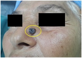
Figure 1: An ulcerated, well-circumscribed and pigmented nodule of the nose.
 Figure 2: Photo of dermoscopy showing an arborizing vessel (blue arrow), chrysalis (green arrow), blue-gray ovoid nests (yellow arrow), and ulceration (white arrow).
The diagnosis of a pigmented Basal Cell Carcinoma (BCC) was initially evoked and the patient was referred for surgical resection. The patient was operated under general anesthesia. She had an excision of the tumor with a sliding flap. The deep re cuts were without abnormalities on the extemporaneous examination.
The postoperative course was simple. There was no recurrence after a follow up of one year.
On microscopic examination, the nodule described macroscopically corresponds to a tumoral proliferation of the dermis arranged in masses in places connected to the epidermis. The tumor cells have a basaloid appearance and reduced cytoplasms sometimes loaded with melanin pigments and are provided with nuclei mono morphs taking a pallisadic disposition at the periphery. The figures of mitoses were present. The stroma was reduced and fibrous. The dendritic melanocytes were also positive for HMB45, and for Melan A (Figure 3). Based on the overall histopathology features, a diagnosis of melanotrichoblastoma was made.
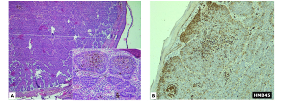
Figure 3: (3A) Nests of basaloid cells within fibromyxoid stroma (H&E x40). Focal pigmentation of basaloid cells (Insert; H&E x100).
(3B) Numerous melanocytes within the nests highlighted by HMB45 immunostaining (original magnification × 100).
(3B) Numerous melanocytes within the nests highlighted by HMB45 immunostaining (original magnification × 100).
DISCUSSION
Trichoblastoma is a rare benign tumor showing differentiation toward the primitive hair follicle [3]. The typical clinical appearance is a solitary, flesh-colored, well-circumscribed, slow-growing tumor that usually progress in size over months to years [4,5]. It can grow up to 3cm. Some cases can reach up to 8-10cm in size [3]. Although they may be present at any age, they most commonly occur in adults in the fifth to seventh decades of life and are equally distributed between males and females. They most often occur on the head and neck with a predilection for the scalp. Although they behave in a benign fashion, cases of malig¬nant trichoblastomas have been reported [5].
Melanotrichoblastoma, also called as pigmented trichoblastoma represents a very rare variant of trichoblastoma in which neoplastic epithelium is colonized by dendritic melanocytic cells [6]. Although melanotrichoblastomas seem to be the most common tumors developing on the nevus sebaceus of Jadassohn , they are usually solitary and may develop in association with other adnexal neoplasms [1]. Due to its relative rarity, its molecular landscape has not been completely understood [6].
According to our knowledge, our case corresponds to the third case of nasal melanotrichoblastoma reported in literature [2,7]. The first case, described by Kamat et al., [2] was reported in a 50-year-old man with a 4.5cm mass over the tip of the nose that slowly enlarged over the course of 6 years. The second case, presented by Sathyaki et al., [7] was described in a 65-year-old Korean woman with a 1cm mass on the right side of the nosethat had been slowly enlarging over the course of 1 year. Comparing these clinical findings with our case demonstrate a relatively similar age of onset in the early to middle-aged adult years. All 3 tumors were slow growing. Additionally, the 3 cases showed well-circumscribed lesions with notable pigmentation.
The differential diagnosis of the pigmented variant includes several epithelial neoplasms with pilar differentiation such as basal cell carcinoma, pigmented pilomatricoma and melanocytic matricoma [6].
Dermocoscopic aspect of trichoblastoma described in the literature is based on small series. We don’t know a specific description for melanotrichoblastoma, given probably the scarcity of this tumor. The presence of ovoid nests, blue-gray cells, and ulceration appears more common in the case of BCC. In our patient, dermoscopy does not differentiate between a melanotrichoblastoma and a BCC [8].
Histologically, pigmented trichoblastomas have distinct melanin pigment at the center of lobules, and dispersed melanocytes between the tumoral cells. These melanocytes show positivity for S100 protein, HMB45, Melan A, and tyrosinase [3]. Epidermal connection, atypia or mitosis is absent. Myxoid stroma and stromal retraction or clefting around basaloid islands, characteristic of basal cell carcinoma, were also absent.
Pigmented trichoblastoma can be a part of a rare syndrome called phacomatosis pigmentokeratotica which is due to mutation of HRAS G123R gene. Treatment consists of surgical resection. No cases of recurrence were noted among the cases reported in the literature [9].
CONCLUSION
This case of melanotrichoblastoma, is being presented because of its rarity and increased chances of clinical misdiagnosis as melanotic neoplasm, nevus and melanoma.
CONFLICT OF INTEREST
The authors declare no conflicts of interest.
REFERENCES
- Kanitakis J, Brutzkus A, Butnaru AC, Claudy A (2002) Melanotrichoblastoma: Immunohisto chemical study of a variant of pigmented trichoblastoma. Am J Dermatopatho 24: 498-501.
- Kamat G, Yelikar B, Shettar S, Karigoudar MH (2009) Pigmented trichoblastoma with sebaceous hyperplasia. Indian J Dermatol Venereol Leprol 75: 506-508.
- Pokharel S, Karki S, Pradhan A, Agrawal S (2017) Melanotrichoblastoma: A rare case report from B. P. Koirala Institute of Health Sciences, Dharan, Nepal. BJHS 2: 145-147.
- Cho HK, Song JS, Kang WH, Ro BI (2009) Pigmented trichoblastoma arising from the nevus sebaceous: A rare case in Korea. Ann Dermatol 21: 406-408.
- Quist EE, DiMaio DJ (2017) Melanotrichoblastoma: A rare pigmented variant of trichoblastoma. Cutis 100: 243-246.
- Saraggi D, Salmaso R, Valentini E, Munari G, Vindigni V, et al. (2017) Pigmented trichoblastoma developed in a sebaceous nevus: HRAS mutation as a common molecular driver. Pathol Res Pract 213: 860-862.
- Sathyaki DC, Riyas M, Roy MS, Swarup RJ, Raghu N (2017) Pigmented Trichoblastoma of Nose: An Unusual Occurrence. J Clin Diagn Res 11: 9-10.
- Hung CT, Chiang CP, Gao CP, Wang WM (2012) Ripple-pattern melanotrichoblastoma arising with in nevus sebaceous. Indian J Dermatol Venereol Leprol 78: 665.
- Li JY, Berger MF, Marghoob A, Bhanot UK, Toyohara JP, et al. (2014) Combined melanocytic and sweat gland neoplasm: Cell subsets harbor an identical HRAS mutation in phacomatosis pigmentokeratotica. J Cutan Pathol 41: 663-671.
Citation: Bouatay R, Soua Y, Jellali S, Moussa, Koubaa J (2019) Melanotrichoblastoma of Nose: A Case Report. J Otolaryng Head Neck Surg 5: 031
Copyright: © 2019 Bouatay R, et al. This is an open-access article distributed under the terms of the Creative Commons Attribution License, which permits unrestricted use, distribution, and reproduction in any medium, provided the original author and source are credited.
Journal Highlights
© 2026, Copyrights Herald Scholarly Open Access. All Rights Reserved!