More than Just a Simple Headache
*Corresponding Author(s):
Diana Henriques PintoDepartment Of Pediatrics, Centro Hospitalar Entre Douro E Vouga, 5 4520-211 Santa Maria Da Feira, Portugal
Email:pinto.dianahenriques@gmail.com
Abstract
A nine-year-old female, without a relevant past medical history, presented in the emergency department with a two-day story of right-sided temporal headache associated with swelling in the same area. No other symptom were reported.
Keywords
Headache; Langerhans cell histiocytosis; Temporal bone
Clinical Image Description
A 9-year-old female, without a relevant past medical history, presented in the emergency department with a two-day story of right-sided temporal headache associated with swelling in the same area. No other symptom were reported. Trauma and recent infectious diseases were denied. On physical examination, she had swelling of right temporoparietal region (Figure 1) with associated cervical lymphadenopathy. The rest of the physical examination was normal.
Blood analysis revealed erythrocyte sedimentation rate of 22mm/hour, but blood count and chemistry were normal. A computed tomography (CT) scan of the head showed a lytic lesion in the right parietal skull, with rupture of the bone cortex (Figure 2).
After two days, she had a magnetic resonance imaging (MRI) that confirmed the suspicion of Langerhans cell histiocytosis (LCH) (Figures 3 and 4).
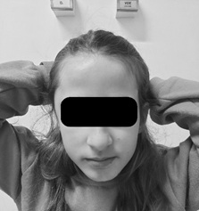 Figure 1: Female children with swelling of right temporoparietal region (arrow).
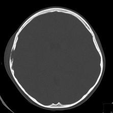 Figure 2: Axial computed tomography image of the head showing a focal lytic lesion of parietotemporal bones, with involvement of soft tissue.
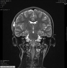 Figure 3: Coronal T2-W MRI of the brain showing parietotemporal lesion, with adjacent soft tissue involvement.
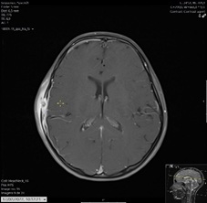 Figure 4: Axial T1-W post-gadolinium show temporal lesion, with adjacent soft tissue involvement.
At staging, positron emission tomography-computed tomography (PET-CT) showed the presence of bone lesions in the right iliac, confirmed by MRI. An iliac biopsy was performed and histology confirmed LCH, classified as multifocal bone, without involvement of risk organs. This child started chemotherapy with prednisolone and vinblastine, according to the LCH-III International Collaborative Treatment Protocol for Children and Adolescents with LCH.
LCH is a rare clonal disease characterized by the abnormal proliferation and accumulation of histiocytic cells in various tissues or organs. These abnormal cells resemble dendritic Langerhans cells but are derived from myeloid dendritic cells, arise from the bone marrow and induce bone resorption, leading to the characteristic lytic bone lesions. It seems that is a result of continuous immune stimulation, but the reason is not completely understood. Most commonly affects bones and skin, but it can also involve the bone marrow, liver, spleen, lungs, pituitary gland, central nervous system (CNS) and other organs. It is accompanied by various degrees of tissue infiltration by inflammatory cells and/or fibrosis [1-4].
LCH may affect any age group but it is considerably more common in children (with peak incidence at 1-3 years of age). A male predominance has been described in some case series. [5] Clinical presentation can be very heterogeneous and so LCH is likely underreported. It can range from unifocal body involvement to disseminated disease with life-threatening complications. Bone involvement is the most common form of presentation in pediatric age (especially bones of the skull).
LCH is classified by the number of organ systems involved (single vs multisystem) and the number of disease sites (uni vs multifocal). Differential diagnostic is very embracing and considerations for multisystemic LCH depends on which organs are affected and thus can be very broad.
The extent of the disease is evaluated with clinical history and physical examination, laboratory studies, imaging and biopsy. CT and/or PET-CT should be done in children over 2 years. Other imaging may be necessary according to the involved sites (for example MRI for brain lesions). Diagnosis is based on histological and immunohistochemical of lesional tissue [6].
The prognosis depends on the form of the disease, its location and response to initial therapy and varies from spontaneous resolution to a progressive multisystemic disorder with organ dysfunction. Among patients with multisystem disease it is important to identify those with CNS (or CNS-risk lesions) and lung and with risk organs (bone marrow, liver, spleen), which affects prognosis and treatment choices.
LCH is a rare disease but it has to be considered in patients with unexplained clinical manifestations of the bone, skin, CNS, lung and liver. An early recognition, diagnosis and multidisciplinary approach is essential and extremely important to improve the outcome.
What Is Known?
#LCH remains a difficult-to-diagnose and has heterogeneous clinical presentation. #MRI and/or CT are important for the diagnosis of LCH.
#Treatment and prognosis depends on the site and extension of the disease.
What is added?
#Pain associated with swelling of the skull should always be investigated.
#Early recognition, diagnosis and multidisciplinary approach can improve the outcome of LCH.
References
- Aubart FC, Ibdaih A, Emile JF, Amoura Z, Abdel-Wahab O, et al. (2021) Histiocytosis and nervous system: from diagnosis to targeted therapies. Neuro-Oncology; 23: 1433-1446.
- William J, Ladner J, Rambie A, Boyer K (2021) Multisystemic Langerhans Cell Histiocytosis in an infant. Radiology Case Reports; 16: 1798-1805.
- Jain A, Kumar S, Aggarwal P, Kumar M, Gupta V (2019) Langerhans cell histiocytosis: An enigmatic disease. The South Asian Journal of Cancer; 8: 183-185.
- Jezierska M, Stefanowicz J, Romanowicz G, Kosiak W (2018) Langerhans cell histiocytosis in children - a disease with many faces. Recent advances in pathogenesis, diagnostic examinations and treatment. Advances in Dermatology and Allergology; 35: 6-17.
- Krooks J, Minkov M, Weatherall A (2018) Langerhans cell histiocytosis in children - History, classification, pathobiology, clinical manifestations and prognosis. Journal of the American Academy of Dermatology; 78: 1035-1044.
- Zhao M, Tang L, Sun S, Fui J (2021) Radiologic findings that aid in the reduction of misdiagnoses of Langerhans cell histiocytosis of the bone: a retrospective study. World Journal of Surgical Oncology; 19: 146.
Citation: Pinto DH, Azevedo I, Costa M, Fraga A, Maia I (2023) More than Just a Simple Headache. J Clin Stud Med Case Rep 10:202
Copyright: © 2023 Diana Henriques Pinto, et al. This is an open-access article distributed under the terms of the Creative Commons Attribution License, which permits unrestricted use, distribution, and reproduction in any medium, provided the original author and source are credited.