Os Calcaneus Secundarius and Calcaneonavicular Coalition - A New Pathophysiological Hypothesis
*Corresponding Author(s):
Vazquez OscarPaediatric Orthopaedics Unit, Geneva Children's Hospital, Geneva University Hospitals, Switzerland
Email:oscar.vazquez@hcuge.ch
Abstract
An Os calcaneus secundarius (OCS) is a rare accessory ossicle of the anterior facet of the calcaneus. The various explanations for its origin involve several contradictory pathophysiological hypotheses, bringing some confusion to the topic. An OCS may be symptomatic or discovered incidentally on X-rays performed for trivial traumas. Some cases have been described in patients presenting with pain mimicking that of a fracture, engendering a peroneal spastic flat foot or associated recurrent sprains. This article analyses four case studies suggestive of OCS and reflects on this condition by reviewing its local anatomy, the biomechanics of the talocalcaneonavicular joint and the various pathophysiological hypotheses concerning it.
Introduction
Finding accessory ossicles around the foot is common; most are asymptomatic, but they can gain clinical relevance through trauma or stress due to the foot’s particularly complex biomechanical system. The Os calcaneus secundarius (OCS) is an accessory ossicle of the anterior facet of the calcaneus, located at the interval between the anteromedial aspect of the calcaneus, the cuboid bone, the talar head and the tarsal navicular [1, 2]. It is rare, with a prevalence of 0.6% to 7%, making it one of the more infrequently found accessory foot bones [3-7]. It is generally bridged to the calcaneus by poorly mobile synchondrosis. The literature reports various explanations for the origin of an OCS, including several contradictory pathophysiological hypotheses, bringing some confusion to the topic. It is, therefore, highly possible probable that several nosological entities presenting in clinically and radiographically similar ways are being confused and conflated. Indeed, an OCS may be discovered incidentally on an X-ray performed for a trivial trauma. However, an OCS may be symptomatic, and cases have been described with patients presenting with pain [8], mimicking a fracture [9], engendering a peroneal spastic flat foot or associated with recurrent sprains [10]. Using the four cases suggestive of OCS that we report here, we reflect on this condition by reviewing the local anatomy, the biomechanics of the talocalcaneonavicular joint and the various pathophysiological hypotheses concerning it.
Case 1
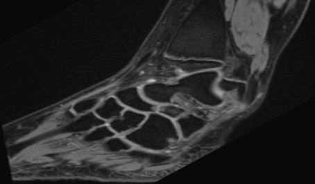
A 16-year-old female, who had presented with repeated left ankle sprains over several years, was consulted at our emergency room for a new left ankle sprain. Radiological evaluation (X-ray and MRI) revealed a discrete oedema of the calcaneal rostrum, with a 4-mm bone fragment between the calcaneum and the navicular bone, making clinicians suspect an avulsion fracture. The talonavicular, spring and bifurcated ligaments were normal. A complementary computed tomography (CT) scan confirmed a 2.8 x 4.5-mm bone fragment with a partially cortical appearance (Figure 1). Treatment was a plaster splint for four weeks. Despite physiotherapy, pain and limited mobility continued, and the patient described pain when walking on non-flat surfaces. On palpation, there was elective pain on the anterolateral side of the mid foot and subtalar motion was restricted. Clinicians proposed a surgical exploration using a lateral approach. We noted synchondrosis/synfibrosis between the calcaneal body and the anterior calcaneal process and between an OCS and the navicular bone. A surgical excision of the OCS was performed, and the extensor digitorum brevis muscle was interposed in the gap left by the removal of the bony piece. At nine months, the patient was walking without pain, had no further sprains and her subtalar motion was no longer restricted.
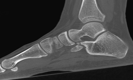 Figure 1: Computed tomography scan image of a partially cortical bone fragment located between the calcaneus and the navicular bone.
Case 2
A 15-year-old female was consulted at our outpatient clinic with a two-year history of multiple right ankle sprains and persisting load-dependent disturbance. She had never presented with local ecchymosis suggestive of a fracture. Radiological evaluation (X-ray and MRI) suggested an OCS but also revealed a synchondrosis/synfibrosis between the tip of the anterior calcaneal process and the navicular bone (Figures 2a-b). On palpation, there was elective pain on the anterolateral side of the mid foot, and she had restricted subtalar motion. Clinicians proposed a surgical exploration using a lateral approach, revealing synchondrosis/synfibrosis between the calcaneal body and the anterior calcaneal process and between an OCS and the navicular bone. A surgical excision of the OCS was performed, and the extensor digitorum brevis muscle was interposed in the gap left by the removal of the bony piece. The treatment quickly and fully resolved the pain and improved subtalar motion, and three months after surgery, the patient resumed her physical activities painlessly.
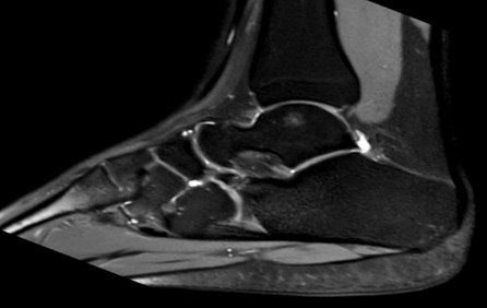
 Figures 2a-b: A sagittal MRI of the right foot suggestive of an OCS but also revealing a synchondrosis/synfibrosis between the tip of the anterior calcaneal process and the navicular bone.
Case 3
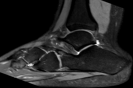
A 13-year-old female attended our outpatient orthopedics clinic with a four-year history of persistent pain after a right ankle sprain. These symptoms precluded her from regular sports activity. On clinical examination, she presented with elective and localized pain on the outer anterolateral side of the foot, and testing of the mid foot revealed subtalar motion restriction without peroneal spasticity. On X-ray and MRI investigation, she was diagnosed with a calcaneonavicular coalition (Figures 3a-b). These examinations also demonstrated a demarcation line located at the base of the anterior calcaneal process, suggesting either an OCS or a stress fracture. Surgical excision was planned and performed using a lateral approach. As in case 2, we noted a synchondrosis/synfibrosis between the calcaneal body and the anterior calcaneal process and between an OCS and the navicular bone. Surgical excision of the OCS was performed laterally, and the extensor digitorum brevis muscle was interposed in the gap left by the removal of the bony piece. Twelve months after surgery, the patient was pain-free and subtalar motion was restored; she could walk without pain and was able to resume her sports activities.
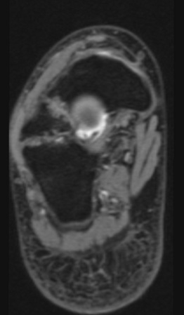 Figures 3a-b: An MRI scan image showing synchondrosis between the OCS and the calcaneus, confirming the calcaneonavicular coalition.
Case 4
A 13-year-old female presented with chronic pain around her right ankle, with many lateral right ankle sprains over the previous three years. Pain was located at the right ankle’s anterolateral aspect, with palpable mass there, and she reported tenderness on palpation of the anterior aspect of the sinus tarsi and around the dorsolateral aspect of the talonavicular joint. Subtalar motion was severely restricted, without hind foot valgus or flat foot deformity. An ossified structure distinct from the distal end of the calcaneus was visible on lateral foot X-ray. Using the 45° oblique view, this bone formation was on top of the anterior process of the calcaneus, superimposed on the calcaneocuboid joint. A CT scan showed a large accessory ossicle (20x12x8mm) attached to the anterior calcaneal facet by a fibro cartilaginous union resembling a synchondrosis. This ossicle was in a space between the anterior aspect of the calcaneus and the navicular bone. Conservative therapy (immobilization with a cast and physiotherapy) was unsuccessful. Surgical treatment consisted of excising this accessory ossicle. After three years of follow-up, the patient was pain-free and her subtalar motion was no longer restricted.
Discussion
Given the heterogeneity of this condition, it seemed important to focus on the different pathophysiological hypotheses concerning it. These gravitate around different concepts involving either the anatomical development of the anterior calcaneal process or the biomechanics of the talocalcaneonavicular joint.
- Developmental Considerations
First, it seems essential to understand how the calcaneum is formed and, above all, how and when the anterior calcaneal process ossifies. The calcaneum’s ossification centre begins to develop by the fourth week of gestational life and becomes visible as a separate structure under the talus on the 55th day. This ossification process is consecutive to intramembranous osteogenesis within the cartilage anlangen [11]. The initiation of calcaneal mineralization follows the invasion of blood vessels and the formation of the periosteal bony collar [12]. This complex ossification centre then develops in different directions centrifugally. It may legitimately be supposed that the anterior calcaneal process, being remote from the calcaneum’s primary ossification centre, remains as a cartilaginous anlage for a relatively long time [13].
- Anatomical considerations
The anterior calcaneal process a saddle-shaped bony promontory is located at the calcaneum’s anterosuperior aspect. The bifurcated ligament is a strong band inserted on its upper surface, dividing at the front in a Y-shaped manner into its calcaneocuboid and calcaneonavicular parts. This ligament is recognized as an important stabilizer of plantar and dorsal ankle flexion, and it is tensioned either by the foot’s inversion and forced plantar flexion [14] or by the eversion of a dorsiflexed foot [3]. Locally, there should be a space between the anterior calcaneal process and the navicular bone, and the calcaneonavicular distance should be greater than 5 mm to prevent any local mechanical impingement [13].
- Biomechanical considerations
The talocalcaneonavicular joint is an arthrodial joint in which the spherical head of the talus is positioned in a concavity formed by the posterior surface of the navicular bone, the anterior articular surface of the calcaneus and the upper surface of the plantar calcaneonavicular ligament. Three principal ligaments are associated with this joint: the dorsal talonavicular ligament, the plantar calcaneonavicular ligament and the calcaneocuboid part of the bifurcate ligament. The calcaneonavicular joint’s biomechanics are thus in dissociable from the talocalcaneonavicular entity, and the whole must be regarded as one functional unit. The talocalcaneonavicular joint generates two types of (gliding and rotating), allowing motion in three axes: inversion/eversion, abduction/adduction and plantarflexion/dorsiflexion. As mentioned above, this functional entity requires a calcaneonavicular clearance greater than 5 mm to prevent any mechanical impingement [13].
- Pathophysiological hypotheses
The pathophysiological hypotheses explaining the occurrence of the OCS have been developed based on these anatomical, developmental and biomechanical considerations. One theory tries to explain its development from a secondary ossification centre at the anterior process of the calcaneus, resulting from a trauma to the immature cartilaginous calcaneus that might have caused the onset of an accessory bone island. In a similar context, other authors hypothesise that the OCS is derived from the failed union of secondary ossification centres: it may be bridged to the calcaneus by poorly mobile synchondrosis, but it can also be completely independent [15].
De Cuveland attributed this structure to a malformation resulting from a persisting inconstant anterior calcaneal process [16]. This hypothesis suggests that there was probably a problem in the chondrified tissue’s process of segmentation into individual anlagen. In Steiner’s concept, the anterior calcaneal process should be considered as an independently developing lateral ray, and he suggested that the OCS represented a persisting supernumerary ray [17]. Niederecker hypothesized that a calcification of the calcaneonavicular part of the ligamentum bifurcatum caused the OCS; however, he provided no explanation for why this calcification occurred [18]. Several authors have evoked a traumatic origin, suggesting that the OCS may derive from fractures of the anterosuperior calcaneal process. These fractures meet the definition of avulsion injuries of the bifurcate ligament caused by the inversion and forced plantar flexion of the foot [14]. Rarely, fractures can be caused by the eversion of a dorsiflexed foot [3]. However, fractures of the anterosuperior calcaneal process can be particularly subtle and easily missed on conventional radiographs. In addition, distinguishing between an OCS and a fracture of the anterior calcaneal process may be very difficult, especially when the fracture is delayed. Using MRI, an ovoid, well-corticated ossicle suggests a diagnosis of OCS, whereas large bony structures with a wide proximal base and adjacent bone marrow oedema suggest a fracture [4]. The clinical reality is somewhat more complex since differentiating between an OCS and pseudarthrosis of a neglected fracture of the anterior calcaneal process can be almost impossible. In both cases, a synfibrosis or synchondrosis may be connecting the calcaneal process to the body of the calcaneus. The main differences between an OCS and pseudarthrosis of the calcaneal anterior process are probably the OCS’s anteromedial positioning in a concave notch at the calcaneal facet and evidence of continuous corticalisation [19]. Finally, several authors have described the OCS as the result of the different developmental stages of a calcaneonavicular coalition [20-22]. In a case involving calcaneonavicular synostosis, one might easily imagine that the mechanical stresses exerted on the coalition could lead to a stress fracture mimicking an OCS.
Our experience, through these four reported case studies, seems to show certain similarities in the radio-clinical presentations, with relatively similar per-operative statuses. Indeed, all four cases presented with synchondrosis or synfibrosis at the calcaneonavicular joint, thus suggesting the incomplete formation of the coalition. Each case presented with a demarcation line at the level of the anterior calcaneal process, with a poorly mobile synchondrosis. We therefore hypothesise that the incomplete coalition noted at the level of the calcaneonavicular space can sometimes lead to an acute or stress fracture at the base of the anterior calcaneal process. Our hypothesis starts with a malformation that disorganizes the talocalcaneonavicular joint by generating a mechanical impingement at the level of the anterior calcaneal process. Since all of our patients suffered from chronic symptoms, a stress fracture seemed to us to be the more plausible cause. However, another explanation might exist in this mechanical context. Indeed, we can imagine that an OCS might be derived from the failed union of secondary ossification centres due to unfavorable mechanical stress. It is impossible to decide between these two hypotheses, even if in our opinion the former seems more coherent.
Conclusion
It remains difficult to affirm whether the Os calcaneus secundarius (OCS) is a true accessory ossicle of the anterior facet of the calcaneus or the result of a fracture of the anterior calcaneal process. Our little case series suggests the occurrence of a stress fracture consecutive to a malformation (calcaneonavicular coalition) that is responsible, in our opinion, for a localized abnormal mechanical load. Finally, the present report emphasizes the importance of a radiographic evaluation, by CT scan or MRI, of the whole calcaneonavicular coalition and any suspect OCS to look out for this particular association.
Conflict of interest statement
The authors declare that they have no conflict of interest.
Funding statement
This study has not received any funding or financial support.
Ethical statement
No request was made to the ethics commission in view of the descriptive nature of the cases and the authorization obtained by the patients.
References
- Vesalius A (1998) On the Fabric of the Human Body: A Translation of De Humani Corporis Fabrica Libri Septem: Book I: The bones and cartilages; Arch Surg; 133: 1251.
- Hohmann G (1948) Fuß und Bein: Ihre Erkrankungen und deren Behandlung. 4th ed. München: Bergmann; 978-3-662-29841-1 (ISBN).
- Hodge JC (1999) Anterior process fracture or calcaneus secundarius: a case report. J Emerg Med. mars; 17: 305-9.
- Mellado JM, Ramos A, Salvadó E, Camins A, Danús M, et al. (2003) Accessory ossicles and sesamoid bones of the ankle and foot: imaging findings, clinical significance and differential diagnosis. Eur Radiol; 13: L164-77.
- Anderson T (1988) Calcaneus secundarius: An osteo-archaeological note. Am J Phys Anthropol; 77: 529-31.
- Keles-Celik N, Kose O, Sekerci R, Aytac G, Turan A, et al. (2017) Accessory Ossicles of the Foot and Ankle: Disorders and a Review of the Literature. Cureus; 9: e1881.
- Krapf D, Krapf S, Wyss C (2015) Calcaneus secundarius – a relevant differential diagnosis in ankle pain: a case report and review of the literature. J Med Case Reports; 9: 127.
- Kepka S, Morel M, Garnier F, Pietra F, Marjanovic N, et al. (2021) A case of an injured calcaneus secundarius in a professional soccer player. BMC Musculoskelet Disord; 22: 374.
- Ersen O, Akyildiz F, Ozyurek S, Sivrioglu AK (2013) Os calcaneus secundarius mimicking fracture. BMJ Case Rep. 2013: bcr2013009954.
- Ceroni D, De Coulon G, Spadola L, De Rosa V, Kaelin A (2006) Calcaneus Secundarius Presenting as Calcaneonavicular Coalition: A Case Report. J Foot Ankle Surg; 45: 25-7.
- Anat J (2001) Ossification in the human calcaneus: a model for spatial bone development and ossification; 199: 609-616.
- Fritsch H, Eggers R (1999) Ossification of the calcaneus in the normal fetal foot and in clubfoot. J Pediatr Orthop; 19: 22-6.
- Lucchesi G, Bonnel F, Wartelle J, Boutry N, Orlando N, et al. (2022) Anatomical characterization of the too-long anterior process of the calcaneum: a computed tomography scan analysis of 69 feet. J Pediatr Orthop B. 32: 47-53.
- Robbins MI, Wilson MG, Sella EJ (1999) MR imaging of anterosuperior calcaneal process fractures. Am J Roentgenol; 172: 475-9.
- Mann RW (1990) Calcaneus secundarius: Description and frequency in six skeletal samples. Am J Phys Anthropol; 81: 17-25.
- De Cuveland E (1966) Zur Herkunft des Os pararticulare am interphalangealen Großzehengelenk. RöFo - Fortschritte Auf Dem Geb Röntgenstrahlen Bildgeb Verfahr; 105: 591-591.
- Der SH (1942) Aufbau des Saugetier-Carpus und Tarsus nach neueren embryologischen Untersuchungen. Rev Suisse Zool; 49: 217-23.
- Niederecker K (1951) Pathologische Veränderungen des Fußskeletts bei Plattfüßen anhand eines größeren Operationsmaterials. Z Orthop; 80: 97-128.
- Hennings R, Voigt P, Kahn T, Josten C, Annette B (2000) Ahrberg Anatomie chirurgicale et radiologique le; 41: 1425-1432.
- Heikel HV (1962) Coalitio Calcaneo-Navicularis and Calcaneus Secundarius: A clinical and radiographic study of twenty-three patients. Acta Orthop Scand; 32: 72-84.
- De Cuveland E (1961) Neue Feststellungen zur Herkunft und Differentialdiagnose inkonstanter Skeletelemente des Fußes. Jahresbericht Borstel; Berlin Heidelberg: Springer; 93-172.
- Leonard MA (1974) The inheritance of tarsal coalition and its relationship to spastic flat foot. J Bone Joint Surg Br; 56B: 520-6.
Citation: Oscar V, Nathaly G, Michael M, Francois C, Dimitri C (2023) Os Calcaneus Secundarius and Calcaneonavicular Coalition - A New Pathophysiological Hypothesis. J Clin Stud Med Case Rep 10:169.
Copyright: © 2023 Vazquez Oscar, et al. This is an open-access article distributed under the terms of the Creative Commons Attribution License, which permits unrestricted use, distribution, and reproduction in any medium, provided the original author and source are credited.