Our Experience with Modified Eloesser Flap in Cases of Postpulmonary Surgery Bronchopleural Fistula with Empyema
*Corresponding Author(s):
Deepi P. AgrawalDepartment Of Cardiovascular And Thoracic Surgery, G.S. Medical And College And KEM Hospital, Acharya Donge Marg, Parel, Mumbai, Maharashtra, 400012, India
Tel:+91 9408514125,
Email:deepi2408@gmail.com
Abstract
Abstract:
Bronchopleural fistula (BPF) is a grave complication of many chronic pulmonary disease and pulmonary resection procedures. Even though the incidence have reduced drastically it has moribund effect but it also comes with multiple therapeutic options. The modified eloesser flap is one of those options. The purpose of our study is to report 4year experience of our institute with MEF.
Methods: A review of 8 patients who had undergone MEF following postpulmonary resection BPF from 2017-2021 was performed.
Results: Adequate drainage was successful in all patients and there were no intraoperative deaths or complications. In all of these patients, the MEF was viable and granulating well or healed.
Conclusion: In the current study, we have confirmed that in a select patient population the MEF is a safe, effective surgical technique for the treatment of advanced empyema with bronchopleural fistula.
Keywords
Bronchopleural Fistula (BPF); Modified Elosser Flap (MEF); Tuberculosis
Introduction
A bronchopleural fistula (BPF) is defined as a communication between a main bronchus or a lobar bronchus and the pleural space after pneumonectomy or lobectomy, with an incidence reported in the literature varying from 0.3 to 20% [1].Despite the recent advances in medicine, Bronchopleural fistula(BPF) remains a debilitating disease process with considerable morbidity and mortality. The most common cause of BPF is infection. Other causes are postpneumenectomy, poor surgical; technique: long bronchial stump, devascularization of bronchus; previous irradiation, malnutrition, concomitant infection, pulmonary tuberculosis, lung abscess, pneumonia [2] The period of hospitalization is long and often multiple operative procedures are necessary for BPF. Sometimes patient are in chronic debilitated state with high risk for GA and thoracoplasty procedure.
Patient And Methods
A retrospective study was performed on the available charts of 8 consecutive patients who underwent the MEF procedure in KEM Hospital, Mumbai from 2017-202. Mean age was 28+10(18-38) with 6 (80%) were male and 2(20%) were female. Out of 8 patients, 5 patients had history of pulmonary tuberculosis and 3 patients had history of fungal infection. Out of 8 patient 3 patient underwent lower lobe lobectomy, 4patient underwent upper lobe lobectomy and 1 patient underwent pneumenectomy. During this period of study, our hospital underwent 75 pulmonary resections, thus the incidence of postresection fistula was 10.6%
Inclusion criteria:
- All the patient with evidence of air leak more than 7 days post pulmonary resection
- All the patient aged 18-45year of age
- Patient who had completed their dose of antitubercular treatment
Exclusion criteria:
- All patient with associated cardiac disease
- All patient with spontaneous bronchopleural fistula
- All patient on active tubercular treatment
Preoperatively, all patients underwent conventional chest roentgenography and standard laboratory evaluation, pulmonary function testing, chest computed tomography, and fiberoptic bronchoscopy scanning were performed in the most all of patients. All patients in this series received various therapeutic interventions before the modified Eloesser procedure. Common therapeutic modalities that were used included bronchoscopic occlusion using plug, glue, tube thoracostomy. Patient’s charts were retrospectively reviewed for age, sex, presenting symptoms, past medical history, past thoracic surgical history, history of ethanol or tobacco use, tuberculosis, fungal infection, nutritional status, preoperative chest roentgenogram findings, white blood cell count (admission and discharge), operative indications, and operative technique. In hospital outcomes included length of stay, days on the ventilator, and morbidity and mortality. Longterm follow-up was attained with a mean follow up period of 1year. All patients underwent the modified Eloesser procedure as described by Symbas and associates [3]. In brief, the patient is turned in a lateral decubitus position, with the involved chest up. An incision is made so as to create an inverted U-shaped flap of skin and subcutaneous tissue over the empyema cavity. The base of the flap is 2 to 4 inches wide, and lies over the most dependent part of the cavity; its length is 2 to 3 inches or equal to the width of two to three ribs and their intercostals spaces. Portions of two or three of the ribs just beneath the U-shaped incision are dissected subperiosteally and removed. The soft tissue portion of the chest wall overlying the abscessed cavity is then resected completing the unroofing of the empyema cavity. After achieving hemostasis and obtaining optimal air seal of the surrounding lung tissue, the U-shaped skin flap is reflected onto the most dependent portion of the abscessed cavity and sutured to the cavity’s floor. The edges of the skin are marsupialized onto the surrounding soft tissue, and a sterile dressing is applied (Figure 1). Postoperative care of the cavity included once-a-day packing with wet-to-dry gauze for approximately 1 months (Figure 2). Thereafter, the cavity can generally be irrigated once daily unless a bronchopleural fistula is present. In which case, continued packing with wet-to-dry gauze is performed secondary to the continued drainage. And once healthy granulating tissue is seen cavity is packed once in 3 days and than simple dressing done (Figure 3).
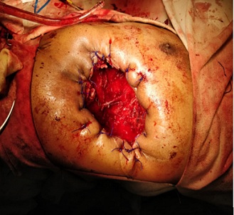 Figure 1: Intraoperative image showing inverted U Shaped Modified Elloser Flap.
 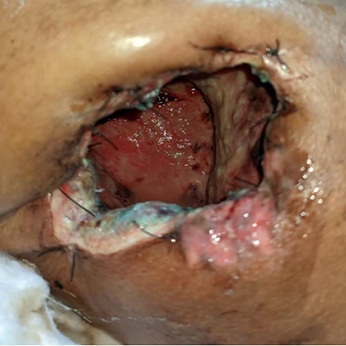 Figure 2: After 1 month of procedure: area of granulation seen.
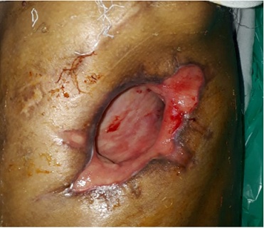 Figure 3: After 3 months of procedure: cavity contracting with healthy granulation tissue.
Results
The most common symptoms of patient undergoing MEF were: dyspnea, fever, tube drainage more than 200ml/day, weight loss, lethargy, chronic productive cough, chest pain, rarely hemoptysis. The presenting white blood cell count for all patients was 18+5 (_1000/_L), whereas the discharge white blood cell count was 8+ 4 (_1000/_L). The most common organisms from preoperative pleural fluid or intraoperative empyema tissue cultures were gram-positive organisms (mainly Staphylococcus and Streptococcus) and gram-negative (mainly Pseudomonas and klebsiella) organisms (Table 1). The most common preoperative interventions were antibiotic therapy (8 patient,100%) tube thoracostomy (8 patients, 100%), bronchoscopic glue (2patient 25%) bronchoscopic plug (2patient, 25%). Intraoperative and inhospital outcomes are presented in (Table 2). Adequate drainage was successful in all patients and there were no intraoperative deaths or complications. There were no death in short term and long term follow up In all of these patients, the MEF was viable and granulating well or healed.
|
Type of microorganism |
Number of patients |
|
Staphylococcus |
2 |
|
Streptococcus |
4 |
|
Klebsiella |
5 |
|
Pseudomonas |
4 |
|
Others: hemophilus, Acinetobacter, etc |
2 |
Table 1: type of microorganisms isolated from patient undergoing MEF
|
Inverted U incision |
8(100%) |
|
Number of ribs resected |
2+1 |
|
Total length of ICU stays |
28+5 days |
|
Total length of postop hospital stays |
100+8 |
|
Total length of overall hospital stays |
150+9 |
Table 2: Intraoperative & Postoperative Outcomes for Patients Undergoing MEF
Discussion
Bronchopleural Fistula is a serious, sometimes fatal disorder which usually follows pulmonary resection but at times occurs spontaneously. It is most commonly associated with tuberculosis, in association with pulmonary resection for other inflammatory dis orders and spontaneously in patients with necrotizing pneumonia, lung abscess, and empyema. Unfortunately, formation of a bronchopleural fistula usually results in long morbid course affecting patient physically as well as psychologically. Floyd, Hollister, and Sealy [4] reported a 28 percent incidence of fistula formation among 32 pneumonectomies and an over-all 10.5 percent incidence among 430 resections carried out for tuberculosis in the early antimicrobial era. But due to current antitubercular drugs and early treatment the rate of fistula formation has decreased, and also the numbers of patients undergoing resection for tuberculosis are twentyfold less than during that period. Technical factors surely play a role, but local preferences for various types of suture material and the use of pleural flaps seem of little consequence. Bronchopleural fistulas occur with all types of bronchial closures including stapling or even after use of intercostals flap or pleural flap. Post surgery fistula may present early, with a fulminating course characterized by sepsis, empyema, purulent sputum, and respiratory insufficiency. Massive air leak from gross disruption of the bronchus requires immediate re exploration and revision of the bronchial closure, initial therapy is open dependent drainage. If a bronchopleural fistula is present, the fistula should be closed by a myoplasty or omentoplasty, followed by single-stage muscle flap closure of the remaining space [5]. Many small fistulas will heal once drainage is established but in cases in which active tuberculosis is present and in other instances wherein the fistula does not close with open drainage and when patient has chronic debilitating factor like malnutrition, weight loss, continuous fever, unable to tolerate General Anesthesia with low lung volume and capacity modified elosser flap seems to be a reasonable approach as compared to thoracoplasty. The Eloesser procedure is considered as a permanent, one-stage procedure and is specifically for those debilitated patients thought not to be a candidate for a muscle flap.
Conclusion
In the current study, we have confirmed that in a select patient population the MEF is a safe, effective surgical technique for the treatment of advanced empyema with bronchopleural fistula. The modified Eloesser procedure remains as an important option in the armamentarium for thoracic surgeons participating in the surgical treatment of chronic, complicated bronchopleural fistula.
References
- Cerfolio RJ (2001) The incidence, etiology, and prevention of postresectional bronchopleural fistula. Semin Thorac Cardiovasc Surg 13: 3-7.
- Magovern CJ, Rusch VW (1994) Parapneumonic and posttraumatic pleural space infections. Chest Surg Clin N Am 4: 561-582.
- Symbas PN, Nugent JT, Abbott OA, Logan WD, Hatcher CR Jr (1971) Nontuberculous pleural empyema in adults. Ann Thorac Surg 12: 69-78.
- Floyd RD, Hollister WF, Sealy WC (1959) Complications in 430 consecutive pulmonary resections for tuberculosis. Surg Gynecol Obstet 109: 467-472.
- 5. Miller JI, Mansour KA, Nahai F, Jurkiewicz MJ, Hatcher CR Jr (1984) Single-stage complete muscle flap closure of the postpneumonectomy empyema space: a new method and possible solution to a disturbing complication. Ann Thorac Surg 38: 227-231.
Citation: Agrawal DP, Jadhav UE (2023) Our experience with modified eloesser flap in cases of postpulmonary surgery bronchopleural fistula with empyema. Archiv Surg S Educ 5: 044.
Copyright: © 2023 Deepi P. Agrawal, et al. This is an open-access article distributed under the terms of the Creative Commons Attribution License, which permits unrestricted use, distribution, and reproduction in any medium, provided the original author and source are credited.