Radiation-Induced Intrapelvic Osteosarcoma Treated with Radical Pelvic Surgery: Case Report and Literature Review
Abstract
Osteosarcoma is a rare but serious sequel of radiotherapy. Surgery used to be the main treatment option in those cases in which in-bloc radical resection was possible. Surgical management of these tumors must take into consideration the possibility of complete resection and surgeries impact the patient’s quality of life. Reconstructive surgery after tumor removal is still controvertial but technically feasible when the tumor is located in the pelvis bone. We present the case of radiation-induced intrapelvic osteosarcoma centered on the right acetabulum with radiological criteria of aggressiveness affecting the hip joint and extending to adjacent soft tissues of the right hemipelvis with suspected internal stop infiltration and isquicoccigeo muscle. Surgery in- bloc resection of the mass performing a total internal hemipelvectomy involving the ilium and the whole of the acetabulum, ischium, pubis and the head of femoral bone were done. Reconstructive pelvic surgery was done with allograft bone inserted in the remainder of the ilium with plates and screws. Multidisciplinary approach to treat pelvic bone sarcoma consist on general surgeon, orthopedic surgeon, plastic surgeon, radiologist and oncologist could be safe and result in good functional results and overall survival.
Keywords
INTRODUCTION
Osteosarcoma is a rare but serious sequel of radiotherapy [1]. Radiation induced osteosarcomas represent about 0.1% of all cancer cases. The latency period can last up to 10 to 14.3 years after treatment [2,3]. The mean time interval between the administration of radiotherapy and the onset of osteosarcoma was of 10 to 14.3 years. Epidemiological data concerning irradiation-induced sarcoma risk showed an average relative risk of 1.42 per 1Gy of dose of gamma irradiation [4].
Surgery used to be the main treatment option in those cases in which in-bloc radical resection was possible. Surgical management of these tumors must take into consideration the possibility of complete resection and surgery´s impact the patient’s quality of life. Reconstructive surgery after tumor removal is still controversial but technically feasible when the tumor is located in the pelvis bone.
We present a case of radiation-induced osteosarcoma of the pelvic bone after radiotherapy for prostate cancer. The diagnosis, treatment and reconstructive surgery approach of irradiation induced intrapelvic bone sarcoma are discussed.
CASE DESCRIPTION
A 65-year-old man was treated in September 2001 with prostatectomy for prostate adenocarcinoma T3aN0M0, ten years later he received radiotherapy treatment due to disease relapse. In June 2014, four years after the completion of treatment with radiotherapy, the patient is referred to our center with a history of lower back pain which radiates to the back of the right leg over the previous four months. X-ray study of the pelvis bone and lumbosacral spine showed degenerative changes in right hip with subchondral sclerosis and marginal osteophytes in the acetabulum. Magnetic resonance revealed a mass centered on the right acetabulum with radiological criteria of aggressiveness affecting the hip joint and extending to adjacent soft tissues of the right hemipelvis with suspected internal stop infiltration and isquicoccigeo muscle (Figure 1).
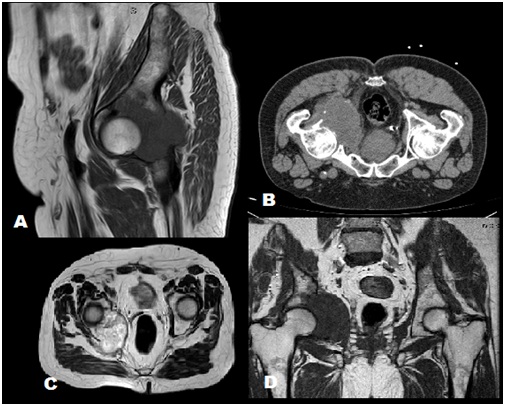 Figure 1: A. MRI sagital view, B. CT scan, C. MRI, D. MRI coronal view.
Computer tomography ruled out any distant metastases. Computer tomography guided needle core biopsy was performed showing the presence of spindle cell proliferation of indeterminate type, suggestive of intermediate-grade spindle cell sarcoma. The tumor cells were characterized by a proliferation of strain mesenchimall growing strands forming solid nests and identifying up to 22 mitotic figures in 10 high power fields. The tumors cells were positive for vimentine, CD 138 and p53 expression. The proliferation cellular index Ki 67 was forty per cent.
A multidisciplinary team consisting of general, orthopedic and plastic surgeons, radiologists and oncologists evaluated the complex case in a committee of tumors. After a joint decision, the patient underwent a selective embolization of the right hypogastric artery and, surgery afterwards.
The patient was positioned in semi lateral position (45°) with a slight elevation of the ipsilateral hip. Utilitarian incision was performed, its ilioinguinal component was advanced medially to the pubic symphysis then, the posterior line is brought to the level of the sacroiliac joint. The abdominal wall musculature, sartorius, and tensor fasciae lata were transected from the iliac crest and reflected away from the ilium. Exposure and mobilization of the femoral vessels, nerve and bladder was performed. Surgery in- bloc resection of the mass performing a total internal hemipelvectomy involving the ilium and the whole of the acetabulum, ischium, pubis and the head of femoral bone were done.
Reconstructive pelvic surgery was done with allograft bone inserted in the remainder of the ilium with plates and screws (Matta Pelvic System. StrykerR). Hip replacement was completed with an acetabular ring (Burch-Schneider reinforcement cage. Zimmer) and proximal femoral prosthesis (MPTM femoral stem. LINKR) A polipropilene mesh served as a reconstructive soft tissue repair (Figure 2).
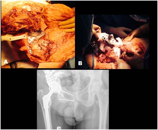 Figure 2: Pelvic reconstruction after tumor resection. 2A and 2B: Surgical procedure images. 2C: Postoperative radiological image.
The postoperative period was uneventful. Total hospital stay was of 38 days. The patient was discharged 35 days after surgery without any complications. At the six months follow-up the patient showed no local recurrence, and the surgical wound had evolved positively. The patient achieved a good functional outcome and recovered a worthy quality of life.
DISCUSSION
In-bloc radical resection could be the main treatment for aggressive pelvic bone sarcoma. Hemipelvectomy may be indicated in the treatment of select tumors of the pelvis and lower extremity. External hemipelvectomy, which involves pelvic resection and amputation of the lower extremity was traditionally been performed in this case. Internal hemipelvectomy represents and alternative approach with reasonable long-term functional outcomes and survival rates [5]. It could involve an extensive bony and soft tissue defects carrying and important reduction of quality of life and functional outcomes.
In this case report we perform a modified internal hemipelvectomy due to the localization of the mass that involves the tree specific regions of the pelvis (ilium, acetabulum and pubis) and our goal was to preserve the lower extremity.
Internal hemipelvectomy used to be the best approach to treat pelvic bone sarcoma due to the best functional outcomes and survival rates, but it is considered a extremely aggressive procedure with a high complication rate. In March 2015 Chao et al. [6] shows a complications rate of 35.1% that is less rate than the previously series that show complications rate of 75% in 1995 by Apffelstaedt [7]. Recently, our group has published his experience in this field [8].
Bone reconstruction is indicated when a complete resection of the ilium is performed that originate a pelvic ring instability. Reconstructive techniques such as iliofemoral artrodhesis, ischiofemoral artrodhesis, allograft or custom-made prosthesis have been published. Reconstruction of bone defect with allograft and a total hip replacement is a surgical treatment with better functional results that other surgical options.
We proposes a multidisciplinary approach that includes orthopedic surgeons and general surgeon to perform hemipelvectomies that includes vascular control, exposure and excision of tumor that is include in the pelvic ring, to reduce complications rates and increase the safety of the surgery improving the patients selection, the elective treatment and the technically approach.
CONCLUSION
Multidisciplinary approach to treat pelvic bone sarcoma consist on general surgeon, orthopedic surgeon, plastic surgeon, radiologist and oncologist could be safe and result in good functional results and overall survival.
CONFLICT OF INTEREST
None of the authors have any conflict of interest to disclose.
FINANCIAL SUPPORT
The manuscript did not receive any funding.
REFERENCES
- Weatherby RP, Dahlin DC, Ivins JC (1981) Postradiation sarcoma of bone: Review of 78 Mayo Clinic cases. Mayo Clin Proc 56: 294-306.
- Iyer R, Jhingran A (2006) Radiation injury: Imaging findings in the chest, abdomen and pelvis after therapeutic radiation. Cancer Imaging 6: 131-139.
- Jemal A, Siegel R, Xu J, Ward E (2010) Cancer statistics, 2010. CA Cancer J Clin 60: 277-300.
- United Nations Scientific Committee on the Effects of Atomic Radiation (2000) Sources and effects of ionizing radiation. Report to the General Assembly with scientific annexes, UNSCEAR, New York, USA.
- Sherman CE, O’Connor MI, Sim FH (2012) Survival, local recurrence, and function after pelvic limb salvage at 23 to 38 years of followup. Clin Orthop Relat Res 470: 712-727.
- Chao AH, Neimanis SA, Chang DW, Lewis VO, Hanasono MM (2015) Reconstruction after internal hemipelvectomy: Outcomes and reconstructive algorithm. Ann Plast Surg 74: 342-349.
- Apffelstaedt JP, Driscoll DL, Karakousis CP (1995) Partial and complete internal hemipelvectomy: complications and long-term follow-up. J Am Coll Surg 181: 43-48.
- Lopez Torres II, Calvo-Haro JA, Mediavilla Santos L, Perez Maanes R, Cuervo Dehesa M, et al. (2018) Post-radiation pelvic sarcomas after radiotherapy treatment of prostate adenocarcinoma. Archieves of Clinical Experimental Surgery 7: 94-99.
Citation: Martín L, Lozano P, Orue-Echebarria MI, Calvo-Haro JA, Cuervo M, et al. (2019) Radiation-Induced Intrapelvic Osteosarcoma Treated with Radical Pelvic Surgery: Case Report and Literature Review. J Surg Curr Trend Innov 3: 014.
Copyright: © 2019 Martín L, et al. This is an open-access article distributed under the terms of the Creative Commons Attribution License, which permits unrestricted use, distribution, and reproduction in any medium, provided the original author and source are credited.