Rare Location of Cavernous Hemangioma
*Corresponding Author(s):
Shirin Hamed-AzzamDepartment Ophthalmology, Baruch Padeh Medical Center, Poriya, Israel
Tel:+972 503636799,
Email:shirinhamedazzam@gmail.com
Abstract
Cavernous hemangioma is a benign vascular tumor. The classic location of orbital cavernous hemangioma is in the intraconal space. The authors present a rare location of cavernous hemangioma in a 47-year-old male. Prior the surgery, it was suspected as fibroma or lipoma. The lesion was surgically excised, and diagnosis of cavernous hemangioma was confirmed by histopathology examination.
BRIEF REPORT
A 47-year-old man who presented with a history of a mass in the right eye brow over 3 months prior to presentation, no history of trauma.
Ocular examination revealed a painless, soft and mobile lesion underneath the tail of the eyebrow. The rest of the examination was unremarkable except of ptosis. Ultrasound scanning found a 3x9mm mass under the lateral eyebrow, suspected for fibroma.
The patient underwent excisional biopsy via lid crease incision (Figure 1A). Histopathology was consistent with cavernous hemangioma (Figure 1B&1C).
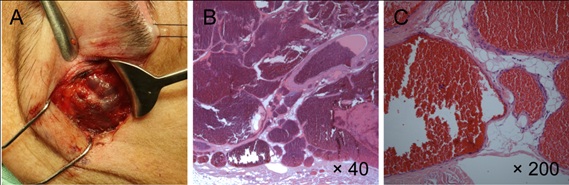 Figure 1: A- intraoperative image of the mass; B,C- Histopathology idenitfied a cluster of densely packed thin-walled blood vessels with wide dilated lumina, which is consistent with cavernous hemnagioma (B,C are magnifications of 40 and 200 respectively).
The most common benign orbital tumor among adults is Cavernous hemangioma and accounts for 3% to 7% of all orbital masses [1]. The typical location is intraconally, often lateral to the optic nerve [2]. Preoperative diagnosis can be made by CT or MRI scans and technetium Tc 99m red blood cell scintigraphic patterns [3].
Cavernous hemangioma of the eyebrow is rare, and should be suspected in patients with extraorbital mass.
REFERENCES
- Shields JA, Bakewell B, Augsburger JJ, Flanagan JC (1984) Classification and incidence of space-occupying lesions of the orbit: a survey of 645 biopsies. Arch Ophthalmol 102: 1606-1611.
- Brackup AH, Haller ML, Danber MM (1980) Hemangioma of the bony orbit. Am J Ophthalmol 90: 258-261.
- Polito E, Burroni L, Pichierri P, Loffredo A, Vattimo A (2005) Technetium Tc 99m-Labeled red blood cells in the preoperative diagnosis of cavernous hemnagioma and other vascular orbital tumors. Arch Ophthalmol 123: 1678-1683.
Citation: Omary R, Mukari A, Jabali-Habib H, Hamed-Azzam S (2020) Rare Location of Cavernous Hemangioma. J Clin Dermatol Ther 6: 044.
Copyright: © 2020 Rawan Omary, et al. This is an open-access article distributed under the terms of the Creative Commons Attribution License, which permits unrestricted use, distribution, and reproduction in any medium, provided the original author and source are credited.