Shear Wave Elasto-Sonography in Gynecological Conditions: State of the Art
*Corresponding Author(s):
Amr Mohamed Abdelhady Sayed AbdelhadyGuy’s And St Thomas’ Hospital, London, United Kingdom
Tel:+44 7888339249,
Email:amr.hady1988@gmail.com
Summary
Shear Wave Elastosonography (SWE) has emerged as a promising, non-invasive diagnostic tool in gynecology, offering quantitative and objective measurements of tissue stiffness. This review article explores the current state of SWE in diagnosing various gynecological conditions, including Polycystic Ovarian Disease (PCOD), uterine adenomyosis, endometrial and subendometrial lesions, uterine fibroids, ovarian cysts, cervical lesions, and pelvic floor disorders. SWE complements conventional ultrasound by providing additional mechanical property assessments, aiding in the differentiation of benign and malignant pathologies. Studies have demonstrated SWE's efficacy in improving diagnostic accuracy for conditions such as PCOD, where increased shear wave velocity (SWV) correlates with ovarian stiffness, and uterine adenomyosis, where SWE offers a cost-effective alternative to MRI. Additionally, SWE has shown potential in evaluating endometrial pathologies, predicting endometrial cancer risk, and assessing treatment responses, such as post-uterine artery embolization in fibroids. Despite its advantages, SWE is not intended to replace traditional imaging but rather to enhance diagnostic precision. The technique is easy to perform, reproducible, and adds minimal time to standard ultrasound examinations. However, further research is recommended to validate its clinical utility across a broader range of gynecological conditions. SWE represents a significant advancement in gynecological imaging, offering a valuable adjunct to conventional diagnostic methods.
Background
Pathological changes in tissue lead to changes in elasticity and stiffness. Strain is deformation of tissue when exposed to pressure. Elasticity is return of tissue to original form when this pressure is removed. Palpation; a clinical method of examination used for thousands of years, is a method to test elasticity and stiffness of tissues. However, palpation is totally subjective and is not suitable for deeper tissues. Elastosonography; a new method to test tissue stiffness. It is suitable for deeper tissue, but it is subjective. Recently, SWE which is objective and quantitative has been developed to test for tissue stiffness.
Introduction
A new promising diagnostic method; elastosonography, is an ultrasound imaging technique that measures tissue strain due to stress applied to it [1]. It could be considered virtual palpation. According to Barr et al., [2], sometimes elastography is called “method of visual palpation”. Palpation has been used in clinical examination for thousands of years; however, it is totally subjective and not suitable for deep organs or tissues. Elastosonography could be considered as a useful diagnostic tool because it is non-invasive, easy to perform, easy to interpret and has a short learning curve towards becoming skilled at the procedure [3]. It will add a few minutes to the time needed for conventional ultrasound.
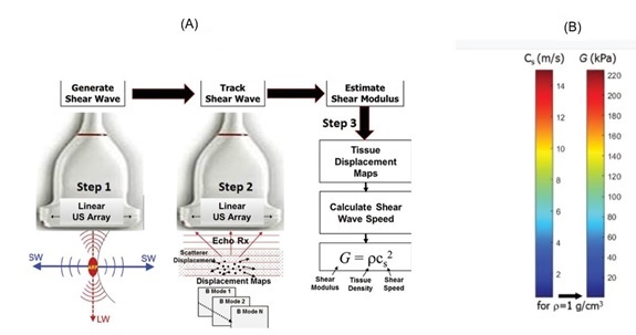
Acoustic Radiation Force Impulse Imaging (ARFI) is a recently developed non-invasive dynamic tissue imaging technique of quantitative elastography [4]. Applying stress to tissue leads to tissue strain (displacement) which is detected and used for obtaining additional information beyond B-mode imaging [5] as shown in figure 1 [6]. When the tissue is harder or stiffer, it strain more (measured in Kpa) and the shear wave moves faster, and its speed (m/s) provides information about tissue hardness [7]. Shear wave elastosonography is becoming an increasingly popular technique in the diagnosis of organs and system diseases [8]. SWE has been used clinically to assess many conditions such as liver diseases [9], thyroid nodules [10], breast cancer [11], preoperative evaluation of axillary lymph nodes status in early breast cancer diagnosis [12], and other disorders.

Figure 1: A) Basic physics of SWE from Taljanovic et al., [6]. In step 1; shear waves are generated using ARF that propagate perpendicular to the primary U/S wave at a lower velocity. In step 2; fast plane wave excitation is used to track displacement and velocity as shear waves propagate, and tissue displacement is calculated using a speckle tracking algorithm. In step 3; tissue displacement is used to calculate shear wave velocity (Cs) and shear modulus (G). (B) Relationship between shear wave velocity and shear modulus expressed as a color bar, which assume es, in this case, tissue density equals that of water (1g/cm3). Actual density estimates will vary for different types of tissue and can also be found using values published in the literature.
Recently, the use of sonoelastography has been reported in the evaluation of endometrium, myometrium, prediction of preterm labor, induction of labor success, pelvic endometriosis and cancer cervix [1,5,11,13]. Hefeda and Zakaria [14] and Wang et al., (2022) [15] suggested that tissue elasticity measured by SWV is considered appropriate for obstetrics and gynecology. SWE should not replace conventional ultrasound imaging, but it is complementary to it [16].
Polycystic Ovarian Disease (PCOD): Conventional U/S examination usually has difficulty presenting the overall morphology and structure of the ovary in patients with PCOS, as physicians can only observe follicles in a limited section of the ovary complicating the process of follicle count [17]. The subjective judgement can easily result in misdiagnosis. Polycystic ovarian morphology can be seen by ultrasound examination in up to 25% of normal ovulating women in whom SWE will not be found increased.
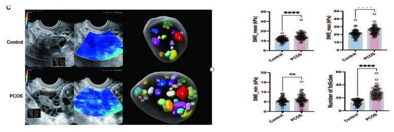
Gursu et al., [4] conducted a study to give insights into the elasticity pattern and SWV in PCOS ovaries compared to ovaries in non-PCOS women. They concluded that easy application of quantitative Sonoelastography Measurements (SWE) provides significant findings about PCOS. They added that the widespread application of SWE in PCOS diagnosis is not a must, but it can be a beneficial diagnostic tool for clinicians. Also, He et al., [17] demonstrated that patients with PCOS (n=59) showed an increased SWV (mean & max) compared to controls (n=56) as shown in figure 2.

Figure 2: Sinus follicle counts and images of Young’s modulus of ovarian medulla in PCOS patients and healthy controls. From He et al., [17].
Uterine Adenomyosis: The most commonly used non-invasive tools to diagnose uterine adenomyosis in clinical practice are: 1) TVS [18], and 2) MRI [19]. According to Acar et al., [20], the subjective nature of B-mode imaging interpretation is a recognized limitation of U/S as a diagnostic modality of this problem and is undoubtedly the cause of wide accuracy range (38.4% to 86.4%). MRI has a good accuracy to diagnose uterine adenomyosis [19]. However, the routine use of MRI is limited by its high cost and less availability. Acar et al., [20] concluded that SWE was accurate to diagnose uterine adenomyosis.
Endometrial and sub endometrial lesions: Endometrial diseases pose a diagnostic challenge due to their nonspecific clinical presentation and features on primary imaging assessment [21]. SWE can assess the mechanical properties of the endometrial lesions and provide a quantitative measure of tissue stiffness and so aids in differentiating different endometrial pathologies [21]. TVS is the first line imaging modality in premenopausal patients with AUB. SWE does not replace conventional TVS, but it is complementary to it [16]. Vora et al., [13] conducted a pilot study to assess the role of SWE in characterizing endometrial and sub endometrial pathologies (endometrial hyperplasia, endometrial polyp, endometrial carcinoma, submucosal leiomyoma and focal adenomyoma). They concluded that SWE is a potential adjunct to ultrasound that provides an additional paradigm to characterize endometrial and sub endometrial lesions.
TV-SWE is an effective additional method of differentiating benign and malignant endometrial lesions when combined with conventional U/S [21].
Uterine fibroid and fibrosarcoma: Samanci and Onal [22] investigated the role of SWE in the evaluation of response to uterine artery embolization (UAE)in patients having uterine leiomyomas. The post UAE 1.5 month stiffness measurements after the procedure were significantly lower than the pre UAE measures. This could be explained by ischemic coagulative necrosis. They concluded that SWE values after UAE, significantly decreased. They added that SWE, with its high reproducibility, could become a useful tool in the follow up of uterine leiomyomas after UAE.
Ovarian cysts: Ciledag et al., [23] conducted a pilot study to determine the appearance of various cystic ovarian lesions on transvaginal real-time ultrasonographic elastography and to investigate its potential in the differential diagnosis of these lesions. They concluded that this diagnostic technique may be used in differentiation of benign and malignant ovarian masses. So unnecessary interventions could be available for benign cysts with solid components. The method used by Ciledag et al., [23] is Strain Elastography (SE) which is subjective qualitative one. Application of the objective quantitative SWE could be more accurate regarding this issue.
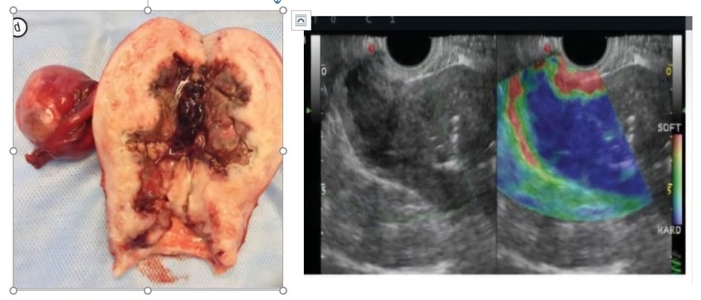
Cervical lesions, figure 3: pathological changes leading to increased stiffness are associated with increased risk of malignancy [24]. Investigations done by Bakay and Golovko, [25] demonstrated that SE included into the ultrasound studies extended the diagnostic possibilities of these methods, allowed to increase the information at examination of cancer process staging. They added that, currently elastography techniques is in the process of development and requires further studies. Fu et al., [26] conducted a study to explore the value of SWE in the differential diagnosis of cervical disease and to evaluate the infiltration of cervical cancer, between October 2014 and January 2017. The mean value of SWS for cervical cancer was significantly higher than that of cervical benign lesions and normal cervix. Also, they concluded that SWE may be used to differentiate between cervical benign lesions and cervical cancer and that SWE is able to evaluate the infiltration of cervical cancer.

Figure 3: Cancer cervix stage IIa stage [26]. Elastography of endophytic tumor which significantly affects the cervix stroma and invades uterine body without leaving its margin. There is a clear border line between dark blue tumor and green colored unchanged stroma. Postoperative specimen confirms absence of invasion outside the uterus.
Perineal bdy (PB): Stress Urinary Incontinence (SUI) is a significant health problem for women, negatively affecting their quality of life [27]. PB, which serves as the anchor of pelvis, is a functional and anatomic complex tissue that maintains urinary continence [28]. The current state of knowledge about correlation of PB and SUI is limited and further studies are needed to understand the role of PB in SUI pathogenesis [29]. The latter authors conducted a study to evaluate the elasticity of PB by SWE in parous women to compare PB elasticity between healthy women and patients with SUI. They found that PB stiffness during Valsalva maneuver in healthy women was higher than in women having SUI.
Pelvic organ prolapse (POP): Assuming that changes of extracellular matrix of connective tissue have specifically contributed to the incidence of POP, it seems reasonable that sonoelastography could be useful tool to evaluate the elasticity of pelvic floor tissue in patients with POP and compare it to those without POP [30]. The latter authors did a pilot study to determine if there are differences in elasticity of the levator ani Muscle (LAM) and vaginal tissue between patients with and without POP. They detected a higher elasticity (lesser stiffness) of the LAM and vaginal tissue in patients with POP than in healthy controls.
Conclusion
Palpation, used in clinical examination for thousands of years is based on detection of changes in elasticity and stiffness of tissue due to pathological conditions. Palpation is subjective and is not suitable for deeper tissues. Elastography, a novel diagnostic modality has been developed to overcome the disadvantages of palpation. Strain elastography was developed first, but it is still subjective and operator dependent. So, SWE has been developed as an objective and quantitative diagnostic modality. It has been studied in many conditions including obstetric and gynecological problems. SWE is easy to perform and interpret, reproducible and add few minutes to the conventional ultrasound examination. More studies are recommended in this area.
Declaration
Author contributions
- All authors participated in writing the paper
- All authors participated in revising and editing the manuscript
Funding statement
This research received no specific grant from any funding agency in the public, commercial, or not-for-profit sectors.
Acknowledgement
The authors would like to thank the reviewers for their valuable comments and suggestions.
Ethical statement
This review article is based on previously conducted studies and does not contain any new studies with human participants or animals performed by any of the authors.
References
- Stoelinga B, Hehenkamp WJ, Brölmann HA, Huirne JA (2014) Real-time elastography for assessment of uterine disorders. Ultrasound Obstet Gynecol 43: 218-226.
- Barr RG, Destounis S, Lackey LB 2nd, Svensson WE, Balleyguier C, et al. (2012) Evaluation of breast lesions using sonographic elasticity imaging: A multicenter trial. J Ultrasound Med 31: 281-287.
- Tessarolo M, Bonino L, Camanni M, Deltetto F (2011) Elastosonography: A possible new tool for diagnosis of adenomyosis? Eur Radiol 21: 1546-1552.
- Gursu T, Cevik H, Desteli GA, Yilmaz B, Bildaci TB, et al. (2021) Diagnostic value of shear wave velocity in polycystic ovarian syndrome. J Ultrason 21: 277-281.
- Bildaci TB, Cevik H, Yilmaz B, Desteli GA (2018) Value of in vitro acoustic radiation force impulse application on uterine adenomyosis. J Med Ultrason (2001) 45: 425-430.
- Taljanovic MS, Gimber LH, Becker GW, Latt LD, Klauser AS, et al. (2017) Shear-Wave Elastography: Basic Physics and Musculoskeletal Applications. Radiographics 37: 855-870.
- Bruno C, Minniti S, Bucci A, Pozzi Mucelli R (2016) ARFI: from basic principles to clinical applications in diffuse chronic disease-a review. Insights Imaging 7: 735-746.
- Cosgrove D, Piscaglia F, Bamber J, Bojunga J, Correas JM, et al. (2013) EFSUMB guidelines and recommendations on the clinical use of ultrasound elastography. Part 2: clinical applications. Ultraschall Med 34: 238-253.
- Hu X, Huang X, Chen H, Zhang T, Hou J, et al. (2019) Diagnostic effect of shear wave elastography imaging for differentiation of malignant liver lesions: A meta-analysis. BMC Gastroenterol 19: 60.
- Aghaghazvini L, Maheronnaghsh R, Soltani A, Rouzrokh P, Chavoshi M (2020) Diagnostic value of shear wave sonoelastography in differentiation of benign from malignant thyroid nodules. Eur J Radiol 126: 108926.
- Zheng X, Huang Y, Liu Y, Wang Y, Mao R, et al. (2020) Shear-Wave Elastography of the Breast: Added Value of a Quality Map in Diagnosis and Prediction of the Biological Characteristics of Breast Cancer. Korean J Radiol 21: 172-180.
- Togawa R, Riedel F, Feisst M, Fastner S, Gomez C, et al. (2024) Shear-wave elastography as a supplementary tool for axillary staging in patients undergoing breast cancer diagnosis. Insights Imaging 15: 196.
- Vora Z, Manchanda S, Sharma R, Das CJ, Hari S, et al. (2022) Transvaginal Shear Wave Elastography for Assessment of Endometrial and Subendometrial Pathologies: A Prospective Pilot Study. J Ultrasound Med 41: 61-70.
- Hefeda MM, Zakaria A (2020) Shear wave velocity by quantitative acoustic radiation force impulse in the placenta of normal and high-risk pregnancy. Egyptian Journal of Radiology and Nuclear Medicine 51: 131.
- Wang XL, Lin S, Lyu GR (2022) Advances in the clinical application of ultrasound elastography in uterine imaging. Insights Imaging 13: 141.
- Ma H, Yang Z, Wang Y, Song H, Zhang F, et al. (2021) The value of shear wave elastography in predicting the risk of endometrial cancer and atypical endometrial hyperplasia. J Ultrasound Med 40: 2441-2448.
- He Y, Deng S, Wang Y, Wang X, Huang Q, et al. (2025) Evaluation of ovarian stiffness and its biological mechanism using shear wave elastography in polycystic ovary syndrome. Sci Rep 15: 585.
- Dueholm M (2006) Transvaginal ultrasound for diagnosis of adenomyosis: A review. Best Pract Res Clin Obstet Gynaecol 20: 569-582.
- Bazot M, Cortez A, Darai E, Rouger J, Chopier J, et al. (2001) Ultrasonography compared with magnetic resonance imaging for the diagnosis of adenomyosis: Correlation with histopathology. Hum Reprod 16: 2427-2433.
- Acar S, Millar E, Mitkova M, Mitkov V (2016) Value of ultrasound shear wave elastography in the diagnosis of adenomyosis. Ultrasound 24: 205-213.
- Soliman BK, Hasan AA, Elfawal Wave MM, Amin MM (2024) Added Value of Transvaginal Real-Time Shear Wave Elastography in the Diagnosis of Endometrial and Sub endometrial lesions. Zagazig University Medical Journal 30: 1718-1727.
- Samanci C, Önal Y (2020) Shearwave elastographic evaluation of uterine leiomyomas after uterine artery embolization: preliminary results. Turk J Med Sci 50: 426-432.
- Ciledag N, Arda K, Aktas E, Aribas BK (2013) A pilot study on real-time transvaginal ultrasonographic elastography of cystic ovarian lesions. Indian J Med Res 137: 1089-1092.
- Bota S, Sporea I, Sirli R, Popescu A, Danila M, et al. (2012) Intra- and interoperator reproducibility of acoustic radiation force impulse (ARFI) elastography--preliminary results. Ultrasound Med Biol 38: 1103-1108.
- Bakay OA, Golovko TS (2015) Use of Elastography for Cervical Cancer Diagnostics. Exp Oncol 37: 139-145.
- Fu B, Zhang H, Song ZW, Lu JX, Wu SH, et al. (2020) Value of shear wave elastography in the diagnosis and evaluation of cervical cancer. Oncol Lett 20: 2232-2238.
- Sasawanga B, Sasawanga N (2013) Stress urinary incontinence in pregnant women: a review of prevalence, pathophysiology, and treatment. Int Urogynecol J 24: 901-912.
- Woodman PJ, Graney DO (2002) Anatomy and physiology of the female perineal body with relevance to obstetrical injury and repair. Clin Anat 15: 321-334.
- Li X, Zhang L, Li Y, Jiang Y, Zhao C, et al. (2024) Assessment of perineal body properties in women with stress urinary incontinence using Transperineal shear wave elastography. Sci Rep 14: 21647.
- García-Mejido JA, García-Jimenez R, Fernández-Conde C, García-Pombo S, Fernández-Palacín F, et al. (2024) The application of Shear Wave Elastography to Determine the Elasticity of the Levator Ani Muscle and Vaginal Tissue in Patients with Pelvic Organ Prolapse. J Ultrasound Med 43: 913-921.
Citation: Abdelhady AMAS, Sayed MA, Soubh SAM (2025) Shear Wave Elasto-Sonography in Gynecological Conditions: State of the Art. HSOA J Reprod Med Gynaecol Obstet 10: 198.
Copyright: © 2025 Amr Mohamed Abdelhady Sayed Abdelhady, et al. This is an open-access article distributed under the terms of the Creative Commons Attribution License, which permits unrestricted use, distribution, and reproduction in any medium, provided the original author and source are credited.

