Journal of Nuclear Medicine Radiology & Radiation Therapy Category: Medical
Type: Opinion Paper
Super Scan in 68Ga-PSMA Ligand PET/CT in Prostate Cancer-Diagnostic Criteria and Its Significance
*Corresponding Author(s):
Manas Kumar SahooDepartment Of Nuclear Medicine And Pet Ct, Medanta-The Medicity, Gurugram, Haryana, India
Tel:+91 9013590865,
Email:drmksahoo@gmail.com
Received Date: Jun 19, 2018
Accepted Date: Aug 07, 2018
Published Date: Aug 21, 2018
Abstract
Superscan in 68Ga-PSMA ligand PET/CT is not very commonly encountered in prostate cancer patients. We present a 70-year-old patient with an elevated PSA serum level of 638 ng/mL after chemotherapy for prostate cancer. 68Ga-PSMA ligand in PET/CT revealed a PSMA over expression of diffuse sclerotic lesions extended to axial and appendicular skeleton. Physiological sites of PSMA over expression such as salivary glands and kidneys were suppressed. This pattern of 68Ga-PSMA PET/CT joined the commonly defined “super-(bone-) scan”. This pattern of appearance in 68Ga-PSMA ligand PET/CT in prostate cancer patients paves way towards targeted therapy like 177Lu-PSMA-617.
Keywords
68Ga-PSMA; PET/CT; Prostate cancer; Superscan
OPINION
Superscan in Bone Scintigraphy (BS) is defined as increased radiotracer uptake in the axial and appendicular skeleton with negligible soft tissue and renal radiotracer uptake [1]. Superscan pattern is also seen in Paget's disease, renal osteodystrophy and non prostatic cancers by such as colon, breast, lung, leukemia, lymphoma, waldenstrom's macroglobulinemia [2].
68Ga-PSMA ligand Positron Emission Tomography (PET/CT) has become an established imaging modality in prostate cancer management [3]. Though unusual, but superscan pattern in 68Ga-PSMA ligand PET/CT has been reported in few cases [4,5].
We observed superscan pattern in a 70-year-old patient with an elevated PSA serum level at 638 ng/mL of a prostate cancer after chemotherapy (Figure 1). Who was referred for 68Ga-PSMA ligand PET/CT for evaluation, which revealed PSMA expressing sclerotic lesions involving almost all the entire skeleton.
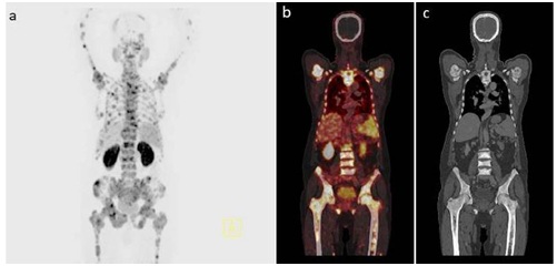
Figure 1: A) Maximum Intensity Projection (MIP) PET image showing increased PSMA expression in the whole skeleton; B) Fused PET/CT image shows increased PSMA expression in multifocal lesions of the appendicular and axial skeleton lesions involving almost all bones of the body with extensive axial as well as appendicular bone involvement; C) Corresponding CT image showing sclerotic lesions involving extensive skeletal sites confirming the metastatic nature of the lesions.
Superscan findings open a new way towards targeted therapy with 177Lu-PSMA-617 for disease control in advanced cases of castration resistant prostate cancers who have progressed after conventional treatments [6,7]. Significant clinical results have so far been achieved with the subsequent use of radiolabeled PSMA ligands in the treatment of CRPC [8].
Superscan in 68Ga-PSMA ligand PET/CT can be defined as increased PSMA expression corresponding with sclerotic/lytic lesions in the CT involving almost the entire skeleton. The background physiological PSMA expression in the salivary glands, gastrointestinal tract should be suppressed. Renal uptake of the radiotracer remains variable and should not be a strict criterion for diagnosis of superscan.
REFERENCES
- Osmond JD 3rd, Pendergrass HP, Potsaid MS (1975) Accuracy of 99mTC-diphosphonate bone scans and roentgenograms in the detection of prostate, breast and lung carcinoma metastases. Am J Roentgenol Radium Ther Nucl Med 125: 972-977.
- Chakraborty PS, Sharma P, Karunanithi S, Bal C, Kumar R (2014) Metastatic superscan on 99mTc-MDP bone scintigraphy in a case of carcinoma colon: Common finding but rare etiology. Indian J Nucl Med 29: 158-159.
- Maurer T, Eiber M, Schwaiger M, Gschwend JE (2016) Current use of PSMA-PET in prostate cancer management. Nat Rev Urol 13: 226-235.
- Lawal I, Vorster M, Boshomane T, Ololade K, Ebenhan T, et al. (2015) Metastatic Prostate Carcinoma Presenting as a Superscan on 68Ga-PSMA PET/CT. Clin Nucl Med 40: 755-756.
- Sager S, Akgün E, Sahin OE, Akgün B, Sönmezoglu K (2017) Superscan Imaging on Ga-68 PSMA PET/CT in Prostate Cancer Patient. J Nucl Med Radiat Ther 8: 320.
- Hofman MS, Violet J, Hicks RJ, Ferdinandus J, Thang SP, et al. (2018) [177Lu]-PSMA-617 radionuclide treatment in patients with metastatic castration-resistant prostate cancer (LuPSMA trial): a single-centre, single-arm, phase 2 study. Lancet Oncol 19: 825-833.
- Aryana K, Zarehparvar Moghadam S, Salek R, Divband G (2018) 177Lu-Prostate-Specific Membrane Antigen Super Scan and Good Response Even After 1 Cycle of Radioligand Therapy. Clin Nucl Med 43: 273-275.
- Virgolini I, Decristoforo C, Haug A, Fanti S, Uprimny C (2018) Current status of theranostics in prostate cancer. Eur J Nucl Med Mol Imaging 45: 471-495.
Citation: Sahoo MK, Shah S (2018) Super Scan in 68Ga-PSMA Ligand PET/CT in Prostate Cancer-Diagnostic Criteria and Its Significance. J Nucl Med Radiol Radiat Ther 3: 010.
Copyright: © 2018 Manas Kumar Sahoo, et al. This is an open-access article distributed under the terms of the Creative Commons Attribution License, which permits unrestricted use, distribution, and reproduction in any medium, provided the original author and source are credited.
Journal Highlights
© 2026, Copyrights Herald Scholarly Open Access. All Rights Reserved!