The Protective Effect of Omega 3 and Vitamin D in Preventing Damage Occurring in Central Nervous System of Neonatal Rats Exposed to Cigarette Smoke in Intrauterine Period
*Corresponding Author(s):
Hayri GözlükgillerDepartment Of Pediatric, Altinbas University, Istanbul, Turkey
Tel:+90 5300780444,
Email:hayrig2000@hotmail.com
Abstract
Purpose: In our study, the toxic effects of cigarette smoke exposure on central nervous system and the protective role of Omega 3 and Vitamin D against these toxic effects were investigated at biochemical and histological level.
Methods and tools: 28 adult female pregnant Wistar-Albino rats were separated into four groups. The first group of rats was our control group. The second group of rats was the group of rats, we have exposed to smoke of 10 cigarettes via puff device 2 hours/day following conception. The third group was the group, to which we have administered Omega 3 (0.5 mg/kg/day) with cigarette smoke, and the fourth group was the group, to which we have administered Vitamin D (42 microgram/kg/day) with cigarette smoke. At the end of the experiment, the sections, collected from brain tissue and medulla spinalis were stained by hematoxylin eosin and .........dye. The levels of Malondialdehyde (MDA) and Fluorescent Oxidation Products (FOU) were the measured biochemical parameters.
Results: We have found the fetal weight and the number of fetuses significantly low only in the group, exposed to cigarette smoke. In the histopathological examination of the brain and spinal cord tissue, we observed that the histopathological changes of the fetuses, exposed to cigarette smoke, were significantly reduced in the group, to which ……Omega 3 and Vitamin D was administered. We have detected that MDA and FOU levels increased with statistical significance in the group, exposed to cigarette smoke, compared to control group (p<0.001), and that MDA and FOU levels were lower in the group, exposed to cigarette smoke along with Omega 3 administration (p<0.001).
Conclusion: We have detected that cigarette smoke caused significant histologic damage in central nervous system tissue of the fetus, and led to oxidative stress, increasing MDA and FOU levels in the tissue. We have found that this damage significantly regressed with Omega 3 and Vitamin D supplements, and that Omega 3 fatty acid is a significant antioxidant, and on the hand Vitamin D has no significant antioxidant effects.
Introduction
Tobacco (cigarettes) is a substance, commonly used today and adversely affecting all systems in the organism due to many toxic agents contained. On the other hand, smoking in pregnancy is an extremely significant issue both in terms of maternal and infant health and in terms of raising healthy generations. Smoking during pregnancy is the most significant preventable risk factor, possibly leading to adverse pregnancy outcomes. In direct proportion to the increased number of cigarettes smoked in pregnancy, complications such as threatened abortion, gestational hypertension, preeclampsia, preterm birth, fetal growth restriction, sudden infant death syndrome, infertility, ablatio placentae, early membrane rupture, and congenital malformations may be observed [1-3].
Smokers are exposed to many chemical compounds such as nicotine, cadmium, lead, and mercury. Cigarette smoke affects the physiological, biochemical, and behavioral functions of central nervous system (CNS). Moreover, exposure to cigarette smoke in pregnancy causes also postnatal morbidity in the form of lower reading and spelling scores and decreased school performance in childhood [4]. In fact, this has been associated even with behavioral disorders in adolescence [5]. Nevertheless, there are studies, performed for its causing psychiatric side effects, attention deficiency, mental retardation and pediatric cancers in children [6,7].
Omega-3 fatty acids are significant fatty acids. They cannot be produced in the body, and they are taken with food. They enter into cell membrane and are present in central nervous system and human brain at high concentrations. They have the ability to cross blood-brain barrier. It has been known for a long time that Omega-3 fatty acids have favorable effects on the cognitive and physical development of the infant in intrauterine period [8-10]. It has been demonstrated that Omega-3 fatty acids are active in nervous system activation, and in development and improvement of memory and cognitive functions, as well as learning capacity [11]. Today it has been included in treatment guidelines for many diseases mainly to help in cardiovascular diseases, in attention deficiency-hyperactivity syndrome, and in enhancing immune system [8,9].
Vitamin D is a significant steroid hormone with a role in calcium hemostasis, known for a long time and with receptors in many different tissues such as nervous system except bone metabolism. Maternal Vitamin D deficiency in pregnancy has been associated with increased risk, related to anomalies in different systems, mainly preeclampsia, gestational diabetes, insulin resistance, low birth weight, preterm labor, and fetal skeletal system [12]. It has been demonstrated in studies conducted that Vitamin D has anti-inflammatory and anti-microbial properties, and plays roles in cell proliferation and differentiation in neurological system, and has neuroprotective effects. Today it has been argued that Vitamin D is classified as a neuro-steroid, and studies on its different functions in brain have been initiated [13].
Although many studies have been conducted in relation to Omega 3 and Vitamin D, studies are insufficient in finding how these vitamins affect fetal central nervous system, exposed to cigarette smoke. More studies are needed to finalize these effects. In our study, we have conducted at biochemical level and with light microscope, the toxic effects of cigarette smoke exposure on central nervous system and the protective role of Omega 3 and Vitamin D against these toxic effects were investigated.
Methods and Tools
28 adult female pregnant Wistar-Albino rats were used in the study. Rats weigh between 150-200 gr. Rats were kept in wire fence mesh cages during the experiment and 7 pregnant rats were kept in each cage. The dimensions of the cages were 30*60*30 cm. Rats were fed with feed (Feed Shop Standard Pellet Rat Feed) and tap water ad libitum in a setting, adjusted for 12 hours of night time and 12 hours of day time. Normal room temperature (22 ± 1°C) and humidity were retained. The pregnancies of rats were confirmed by solid, yellow vaginal plaque formation. The appearance of vaginal plaque was accepted as the first day of the pregnancy.
Our first group of rats was our control group. It was ensured that rats got pregnant after mating. Mother rats were spontaneously observed without any procedure. Following birth, baby fetuses were kept in the same cage with their mothers, and they were fed only with breast milk. We have exposed the second group of rats to smoke of 10 cigarettes via puff device 2 hours/day following conception. Also we similarly exposed the neonatal fetuses to cigarette smoke 2 hours/day. Cigarette smoke 2 hours/day + Omega 3 (0.5 mg/kg/day) were administered to the third group of pregnant rats following conception. We kept the neonatal fetuses in the same setting with the mother for 10 days, and we ensured that they received only breast milk and they were exposed to cigarette smoke. Cigarette smoke 2 hours/day + Vitamin D (42 microgram/kg/day) were administered to the fourth group of pregnant rats. We kept the neonatal fetuses in the same setting with the mother for 10 days, and we ensured that they received only breast milk and they were exposed to cigarette smoke. We administered Vitamin D and Omega 3 intraperitoneally.
Baby fetuses were examined macroscopically to determine whether there is any anomaly or pathology and their weights were recorded. On Day 10 after the birth, baby fetuses were administered ketamine for anesthesia and their brain and spinal cord tissue were collected by appropriate incision to be sent to pathology and biochemistry departments. Routine histological tissue tracking and paraffin blocks were applied to these tissues. Sections of 5- 6µm thickness, prepared from paraffin blocks, were stained with hematoxylin eosin, Caspase- 3 and neuro-filament. Apoptosis was assessed immunohistochemically with Caspase-3 and degeneration and demyelination were assessed in nerve tissue with neuro-filament staining [7]. Brain and spinal cord tissue preparations were selected from each group with systematic random sampling method.
Histopathological Scoring
Demyelination: Absent 1 point, presence of partial demyelized areas 2 points, extensive demyelized areas 3 points.
Degeneration: Absent 1 point, partial degeneration 2 points, extensive degeneration 3 points.
Apoptosis: Absent 1 point, partial apoptosis 2 points, extensive apoptosis 3 points cohesion: Absent 1 point, partial cohesion 2 points, extensive cohesion 3 points.
The levels of Malondialdehyde (MDA) and Fluorescent Oxidation Products (FOU) were the biochemical parameters, measured in brain and spinal cord tissue to determine whether there are differences among the groups in terms of oxidation products. MDA levels were measured using the fluorometric method as described by Wasowicz. Fluorescent oxidation products were measured with modified Shimasaki methods. Briefly tissue homogenates were extracted with ethanol-ether (3/1 v/v) and were measured by spectroflurometrically at a wavelength of 360/430 (excitation/emission wavelength).
Statistical Analysis
Whether the distributions of continuous variables were normally or not was determined by Shapiro-Wilk test. Levene test was used for the evaluation of homogeneity of variances. Data were expressed as mean ± SD or median (interquartile range), where applicable. While, the mean differences among groups were analyzed by One-Way ANOVA, otherwise, the Kruskal Wallis test was applied for the comparisons of not normally distributed data. When the p value from One-Way ANOVA or Kruskal Wallis test statistics were statistically significant, post-hoc Tukey HSD or Conover’s multiple comparison test were used to know which group differs from others. Data analysis was performed by using IBM SPSS Statistics version 17.0 software (IBM Corporation, Armonk, NY, USA). A p value less than 0.05 was considered as statistically significant.
Results
In the analysis, performed in relation to the number of fetuses, obtained from the groups, the difference among the groups was found to be statistically significant. We observed that the number of fetuses in the group, exposed to cigarette smoke, decreased significantly (p < 0.001). We observed that the number of fetuses in the group, to which Omega 3 and Vitamin D were administered, increased, and this number was similar to the number of fetuses in the control group. We found that there was a statistically significant difference among the groups in terms of mean body weight (p < 0.001), and that fetus weights were significantly low only in the group, exposed to cigarette smoke. Fetus weights were found to be similar in the control group and in the group, to which Omega 3 and Vitamin D were administered. We assessed fetuses in terms of head, body, and extremity anomalies, and we did not find any significant difference among the groups. On the other hand, in the histopathological examination of the brain and spinal cord tissue, ......we observed that MDA and FOU levels, used as significant parameters in revealing oxidative damage, increased with statistical significance in the group, exposed to cigarette smoke, compared to the control group (p < 0.001), and we detected that MDA and FOU levels were lower in the group, exposed to cigarette smoke along with Omega 3 administration (p < 0.001). On the other hand, MDA and FOU levels were observed to be low in the group, to which Vitamin D was administered, but we did not find any statistical significance.
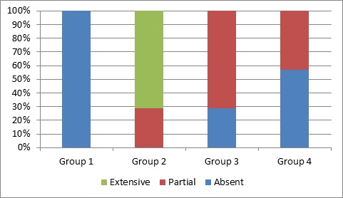
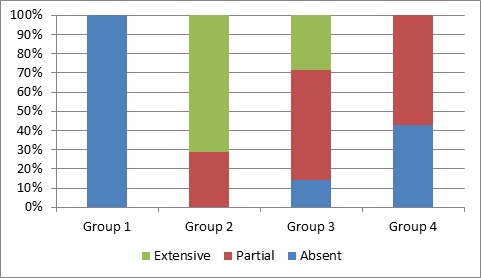
Statistically significant difference was detected among the groups in terms of demyelination scores (p < 0.001). The demyelination scores of the Cigarette (Group 2), Omega-3 (Group 3), and Vitamin D (Group 4) groups were higher with statistical significance compared to Control group (Group 1), respectively (p < 0.001, p < 0.001, and p=0.030). The demyelination scores of Omega-3 and Vitamin D groups were lower with statistical significance compared to the group, exposed to cigarette, respectively (p < 0.001 and p < 0.001). No statistically significant difference was observed among Omega-3 and Vitamin D groups (p=0.137) (Table 1 & Figure 1). Statistically significant difference was detected among the groups in terms of degeneration scores (p < 0.001). The degeneration scores of the Cigarette (Group 2), Omega-3 (Group 3), and Vitamin D (Group 4) groups were higher with statistical significance compared to Control group (Group 1), respectively (p < 0.001, p < 0.001, and p=0.010). The degeneration scores of Omega-3 and Vitamin D groups were lower with statistical significance compared to the group, exposed to cigarette, respectively (p=0.024 and p < 0.001). The degeneration score of Vitamin D group was also lower with statistical significance compared to Omega-3 group (p=0.018) (Table 1 & Figure 2).
|
|
Group 1 |
Group 2 |
Group 3 |
Group 4 |
p-value † |
|
Demyelination |
|
|
|
|
< 0.001 |
|
Absent |
7 (100.0%)a,b,c |
- |
2 (28.6%) |
4 (57.1%)c,e |
|
|
Partial |
- |
2 (28.6%) |
5 (71.4%)b,d |
3 (42.9%) |
|
|
Extensive |
- |
5 (71.4%)a,d,e |
- |
- |
|
|
Degeneration |
|
|
|
|
< 0.001 |
|
Absent |
7 (100.0%)a,b,c |
- |
1 (14.3%) |
3 (42.9%) |
|
|
Partial |
- |
2 (28.6%) |
4 (57.1%)b,d,f |
4 (57.1%)c,e,f |
|
|
Extensive |
- |
5 (71.4%)a,d,e |
2 (28.6%) |
- |
|
|
Apoptosis |
|
|
|
|
< 0.001 |
|
Absent |
7 (100.0%)a,b |
- |
4 (57.1%)b,d |
6 (85.7%)e |
|
|
Partial |
- |
2 (28.6%) |
3 (42.9%) |
1 (14.3%) |
|
|
Extensive |
- |
5 (71.4%)a,d,e |
- |
- |
|
|
Cohesion |
|
|
|
|
< 0.001 |
|
Absent |
7 (100.0%)a,b,c |
- |
4 (57.1%)b,d |
4 (57.1%)c,e |
|
|
Partial |
- |
1 (14.3%) |
3 (42.9%) |
3 (42.9%) |
|
|
Extensive |
- |
6 (85.7%)a,d,e |
- |
- |
|
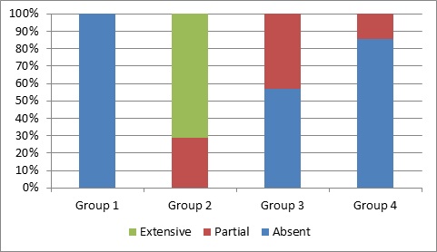
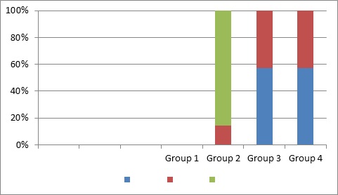
Table 1: Frequency distributions regarding histopathological evaluation.
Note: Data were shown as number of cases and (percentage), † Kruskal Wallis test, a: Group 1 vs Group 2 (p < 0.001), b: Group 1 vs Group 3 (p < 0.05), c: Group 1 vs Group 4 (p < 0.05), d: Group 2 vs Group 3 (p < 0.05), e: Group 2 vs Group 4 (p < 0.001), f: Group 3 vs Group 4 (p=0.018).
 Figure 1:
|
Group 1 |
Group 2 |
Group 3 |
Group 4 |
|
|
Absent |
100 |
0 |
28.6 |
57.1 |
|
Partial |
0 |
28.6 |
71.4 |
42.9 |
|
Extensive |
0 |
71.4 |
0 |
0 |
 Figure 2:
|
Group 1 |
Group 2 |
Group 3 |
Group 4 |
|
|
Absent |
100 |
0 |
14.3 |
42.9 |
|
Partial |
0 |
28.6 |
57.1 |
57.1 |
|
Extensive |
0 |
71.4 |
28.6 |
0 |
Statistically significant difference was detected among the groups in terms of apoptosis scores (p < 0.001). The apoptosis scores of Cigarette (Group 2) and Omega-3 (Group 3) groups were higher with statistical significance compared to Control group (Group 1), respectively (p < 0.001 and p = 0.030). The apoptosis scores of Omega-3 and Vitamin D groups were lower with statistical significance compared to the group, exposed to cigarette, respectively (p < 0.001 and p < 0.001). No statistically significant difference was observed among Control group and Vitamin D group, and Omega-3 group and Vitamin D group (p=0.450 and p=0.137) (Table 1 & Figure 3).
 Figure 3:
|
Group 1 |
Group 2 |
Group 3 |
Group 4 |
|
|
Absent |
100 |
0 |
57.1 |
85.7 |
|
Partial |
0 |
28.6 |
42.9 |
14.3 |
|
Extensive |
0 |
71.4 |
0 |
0 |
Statistically significant difference was detected among the groups in terms of cohesion scores (p < 0.001). The cohesion scores of the Cigarette (Group 2), Omega-3 (Group 3), and Vitamin D (Group 4) groups were higher with statistical significance compared to Control group (Group 1), respectively (p < 0.001, p=0.037, and p=0.037). The cohesion scores of Omega-3 and Vitamin D groups were lower with statistical significance compared to the group, exposed to cigarette, respectively (p < 0.001, and p < 0.001). No statistically significant difference was observed among Omega-3 and Vitamin D groups (p=1.000) (Table 1 & Figure 4).
 Figure 4:
|
Group 1 |
Group 2 |
Group 3 |
Group 4 |
|
|
Absent |
0 |
57.1 |
57.1 |
|
|
Partial |
0 |
14.3 |
42.9 |
42.9 |
|
Extensive |
0 |
85.7 |
0 |
0 |
Discussion
It is known that smoking causes changes in histological structure and functions of brain tissues. Abreu-Villaca, have indicated that smoking caused cell damage during fetal development, and prevented synaptic maturation and intercellular communication. Moreover, they have stated that nicotine demonstrated neurotoxic effect for brains at developmental age. Slotkin, have reported that nicotine administration led to neuron damage in cerebral cortex of adolescent rodents. In this study, we have conducted, vacuolization at molecular layer and neuroglial cell infiltration were observed in prefrontal cortex of rats, exposed to cigarette smoke. Moreover, sporadic pyknotic cells were detected. In conclusion, the findings, we have obtained in relation to cigarette exposure, show parallelism with the studies above. In these studies, previously conducted, an increase in Malondialdehyde (MDA) levels, which is an indicator of lipid peroxidation and a decrease in Superoxide Dismutase (SOD) and Glutathione Peroxidase (GSH-Px) values, which are antioxidant enzymes, were detected in lung, liver, and testis tissues of rats, exposed to cigarette smoke. It has been indicated that these disorders may be due to oxidative damage, occurring via cigarette exposure. Brain is sensitive to oxidative stress, since its rate of oxygen consumption is high. Additionally, it has been reported that the antioxidant defense system is weak in the brain, and that smoking leads to oxidative stress also in these tissues.
In experimental studies, it has been detected that post-trauma inflammation decreased after the administration of 1,25(OH)2 D3 and progesterone to rats with traumatic brain damage, and it has been confirmed that they had neuroprotective effects. Another mechanism, accepted in explaining the neuroprotective effect of Vitamin D, is Vitamin D’s reducing the level of Reactive Oxygen Substrates (ROS). It has been indicated that 1,25(OH)2D3 increased antioxidant effect in glia and neurons, and decreased ROS in dead cells. It is argued that Vitamin D cannot be regarded only as a vitamin and that it plays roles in many mechanisms also in the brain, and therefore, its level is closely associated with the development of some neurological diseases. It is asserted that Vitamin D deficiency may lead to adverse effects in the brain at various stages of life, and may cause Parkinson’s disease, Alzheimer’s disease, MS, ALS, etc. It has been suggested in relation to Vitamin D metabolism in the brain that the neuroprotective effect of CY- Vitamin D, one of cytochrome P450 enzyme systems, in glial cells, may be demonstrated by decreased expression of L-type calcium channels or by increased VDR level. Therefore, vitamin D deficiency can be regarded as a factor, increasing the risk of formation of neurological diseases. The increased VDR expression in embryological period is suggested to increase apoptosis, and to decrease mitosis, and to affect cell proliferation, and may play a significant role also in neuron development. However, 1,25(OH)2D3 activity in the brain cannot be fully understood yet. It is considered that 1,25(OH)2D3 reduces proliferation in neuroblastoma, and also the active mechanism herein when cellular differentiation increases is the conversion of Vitamin D to its inactive metabolites, decreasing proliferation and ensuring a balance. It is argued that 1,25(OH)2D3 has a strong regulatory effect in signal transduction of Neuron Growth Factor (NGF), which is especially apparent in developing neurons, and therefore, may be important in the development and the migration of neurons in the brain.
References
- Ananth CV, Savitz DA, Luther ER (1996) Maternal cigarette smoking as a risk factor for placental abruption, placenta previa, and uterine bleeding in pregnancy. Am J Epidemiol 144: 881-889.
- Haustein KO (1999) Cigarette smoking, nicotine and pregnancy. Int J Clin Pharmacol Ther 37: 417-427.
- Wollmann HA (1998) Intrauterine growth restriction: Definition and etiology. Horm Res 49: 1-6.
- Gilman SE, Gardener H, Buka SL (2008) Maternal smoking during pregnancy and children's cognitive and physical development: A causal risk factor? Am J Epidemiol 168: 522-531.
- Keyes KM, Keyes MA, March D, Susser E (2011) Levels of risk: Maternal-, middle childhood-, and neighborhood-level predictors of adolescent dis-inhibitory behaviors from a longitudinal birth cohort in the United States. MentHealth Subst Use 4: 22-37.
- Batstra L, Hadders-Algra M, Neeleman J (2003) Effect of antenatal exposure to maternal smoking on behavioural problems and academic achievement in childhood: Prospective evidence from a Dutch birth cohort. Early Hum Dev 75: 21-33.
- Hofhuis W, Merkus PJ, de Jongste JC (2002) Negative effects of passive smoking on the (unborn) child. Ned Tijdschr Geneeskd 146: 356-359.
- Young G, Conquer J (2005) Omega-3 fatty acids and neurophysichyatric Reprod Nutr Dev 45: 1-28.
- Balk EM, Lichtenstein AH, Chung M, Kupelnick B, Chew P, et al. (2006) Effects of omega-3 fatty acids on serum markers of cardiovascular disease risk: A systematic review. Atherosclerosis 189: 19-30.
- Guest J, Garg M, Bilgin A, Grant R (2013) Relationship between central and peripheral fatty acids in humans. Lipids Health Dis 12: 79.
- Konagai C, Yanagimoto K, Hayamizu K, Han L, Tsuji T, et al. (2013) Effects of krill oil containing n-3 polyunsaturated fatty acids in phospholipid form on human brain function: A randomized controlled trial in healthy elderly volunteers. Clin Interv Aging 8: 1247-1257.
- Gürz AA, Igde FA, Dikici MF (2015) Fetal and maternal effects of vitamin D. Konuralp Tlp Dergisi 7: 69-75.
- Eyles DW, Burne TH, McGrath JJ (2013) Vitamin D, effects on brain development, adult brain function and the links between low levels of vitamin D and neuropsychiatric disease. Front Neuroendocrinol 34: 47-64.
Citation: Gözlükgiller H (2021) The Protective Effect of Omega 3 and Vitamin D in Preventing Damage Occurring in Central Nervous System of Neonatal Rats Exposed to Cigarette Smoke in Intrauterine Period. J Neonatol Clin Pediatr 8: 085.
Copyright: © 2021 Hayri Gözlükgiller, et al. This is an open-access article distributed under the terms of the Creative Commons Attribution License, which permits unrestricted use, distribution, and reproduction in any medium, provided the original author and source are credited.

