Journal of Animal Research & Veterinary Science Category: Agriculture
Type: Research Article
Anatomical Study on the Pelvic Diaphragm of Male Balady Dog
*Corresponding Author(s):
Mohammed A NazihLecturer Of Anatomy And Embryology, Faculty Of Veterinary Medicine, New Valley University, New Valley Governorate, Egypt
Tel: +20 1003732616,
Email:anatomynazih@yahoo.com
Received Date: Jun 24, 2019
Accepted Date: Jul 18, 2019
Published Date: Jul 25, 2019
Abstract
The study was applied on 13 apparently healthy male balady dogs, obtained from the laboratories of faculty of veterinary medicine - new valley university. The specimens were carefully dissected in order to study the anatomical structural characteristics of the pelvic diaphragm. The latter, guarded the pelvic outlet in two main triangular areas; anal and urogenital. The diaphragm represented in three parts; dorsal, intermediate and ventral. The former was the ischiorectal and composed of the coccygeus muscle group and the anal part. The intermediate division included the perineal body and muscle and the ventral one was the urogenital diaphragm. The perineal fasciae were declared superficial and deep and the membranous layer of the former was the colle’s fascia that inclosed the superficial perineal pouch. The deep perineal fascia represented the urogenital diaphragm and the deep perineal pouch was formed by the reflected perineal membrane on the ischiurethralis muscle. Some anatomical differences were noticed and discussed with that of human anatomy. The study was a guide to understand the anatomical characteristics of the clinically significant area of the most frequent perineal hernia.
Keywords
Anatomy; Balady; Dog; Pelvic diaphragm
INTRODUCTION
Dog’s health and reproductive quality, represented a valuable economical importance for owners as well as saving the breed Wells [1] Friedmann and Son [2]. Studding the pelvic diaphragm among dogs, triggered the attention of previous authors Sluijs & Sjollema [3], Moraes et al., [4] and Sprada et al., [5]. Regarding its role in frequently recognized clinical incidence of the perineal hernia, the weakness of the perineal muscles and fasciae were the main causes of the perineal herniation Ferreira & Delgado [6] and Costa Neto et al., [7]. The formers and Hedlund [8], Bellenger and Canfield [9] and Ribeiro [10] added that the size of prostate gland affected the pelvic diaphragm integrity in male dogs. Hall et al. [11] in dog compared the anatomical homologies of the perineal region in male and female dogs. Anatomically, the pelvic diaphragm consisted of the coccygeus muscle, levator ani muscle, external anal muscle, internal obturator muscle and sacrotuberous ligament Bellenger and Canfield [9]. In regards to the limited available literatures, the present work aimed to focus on the morphological characteristic features of the pelvic diaphragm in balady male dog.
MATERIAL AND METHODS
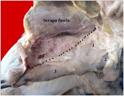
Thirteen adult apparently healthy male balady dogs were collected from stray and laboratories of the faculty of Veterinary Medicine New Valley University. The dogs were slaughtered and fixed by formalin 10% solution for four days. The pelvic cavity with connected hind limbs was cut out from the preserved specimens. Two anatomical regions were intended and prepared for the study; the ischiorectal fossa and the urogenital triangle figure 1. For the former aspect, the dissection started by an upward fixation of the tail and incising the skin at the median longitudinal line ventrally to the root of the tail reaching the dorsal border of the anus. Another circular one surrounded the latter and extended ventrally to the level of ischial tuberosities where a horizontal cut was applied to the point of the tuberosties. Carefully the skin was reflected for dissecting the superficial aspect of the ischiorectal fossa.
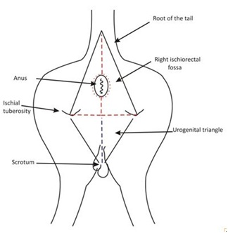
Figure 1: A diagram showing the hind limbs of male dog (caudal view) demonstrate the lines of the anatomical incisions.
The red dotted lines for ischiorectal fossae and the bluish one for the urogenital triangle.
For examining the urogenital triangle, a median longitudinal incision was cut along the scrotum and prepuce to the level of the pubic symphysis. All dimensions were measured using varineir caliper.
It is interesting to note that, there was a thick fatty coat covered the areas of dissection, so carful dissection is preferred.
RESULTS
The pelvic outlet is bounded caudally by a bilateral fasciomuscular closure, the pelvic diaphragm. It supports the retroperitoneal pelvic organs to prevent their perineal prolapse. The perineum extends deeply to the level of imaginary lines from the apex of the sacrum dorsally, ischial arch and tuberosities ventrally and sacroischiatic ligament laterally, as well as superficially along the region which extends ventrally from the anus to the level of the caudal end of pelvic symphysis. It consists of two anatomical triangular areas; dorsal and ventral, anal and urogenital respectively figure 2. The anal triangle is the area that is included in the lines between the sacral apex dorsally, sacroischiatic ligament laterally and ischial tuberosities ventrally. The region is composed of a bilateral ischiorectal fossae, each of them extends from the sacral apex dorsally, anal opening ventromedially, ischial tuberosities ventrolaterally and sacroischiatic ligament laterally figure 3a. The urogenital triangle extends ventrally to the former and its base facing dorsally between the ischial tuberosities while its apex reaches the level of the pubic symphysis. The pelvic diaphragm lodges in the deep and superficial aspects of the perineum and is categorized in three parts; dorsal, intermediate and ventral. The former, the ischiorectal part includes the coccygeus muscle group and the anal part which forms the suspensory apparatus of the anus. The latter, consists of the rectococcygeus and the retractor penis muscle.
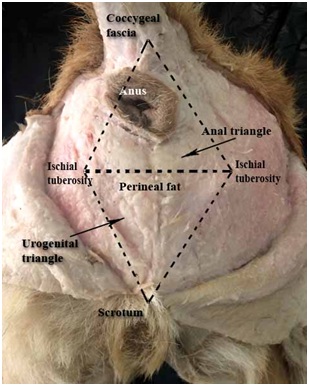
Figure 2: A photograph showing the areas of dissection (caudal view).
The intermediate perineal part represents the perineal body and the longitudinal cutaneous perineal muscle and the ventral one is composed of the urogenital diaphragm. The perineal fasciae represent the latter and is composed of superficial and deep parts.
The dorsal part
The Ischiorectal part (Coccygeus muscle group) comprises the lateral coccygeus muscle and the medial coccygeus group (corresponding to levator ani muscle). The lateral coccygeus muscle (Figures 3a,3b,4-7) is a thin flat quadrilateral in shape, arising along the ischiatic spine and its fibers directed caudo- dorsally and medially. The muscle is inserted in the lateral aspects of the first three caudal vertebrae. It guards the caudolateral aspect of the sacroischatic angle and is bounded laterally by the ischiorectal fat bad and sacrotuberous ligament, while medially it is related to the medial coccygeus group. The muscle has ventral, cranial, dorsal and caudal borders; the former, measures about 1.8-2 cm length, 2.7-2.9cm for the cranial, 3.6-3.8cm for the dorsal and 4.5-4.7cm for the caudal one. The medial coccygeus muscle group (Levator ani muscle) (Figures 3a,3b,4,7-10,11a,11b) (3a,3b,4,is the main closure of the pelvic outlet and is considered the chief muscle of the pelvic diaphragm. The group consists of iliococcygeus, pubococcygeus and ischiococcygeus muscle. The first, (Figures 8-10) is a flat triangular muscle attached from the cranial border of the ilio-pubic junction and its fibers directed caudo- dorsally and medially to be inserted in the lateral aspect of 4th caudal vertebrae. Its base facing ventrally and measures about 2.1-2.3cm, 5.4-5.6 cm for the cranial border as well as 4.6-4.8 cm for the caudal one. The muscle is related laterally to the shaft of ilium and medially to the pubococcygeus muscle and obturator nerve. It overlaps the craniolateral aspect of the latter muscle, where its caudal fibers join that of pubococcygeus forming single muscle bundle. The pubococcygeus muscle (Figures 3a,3b,4,7-10) is the largest part of the group, the thin and flat muscle fibers arise from the dorsal and lateral aspect of the pubis and pelvic symphysis. The muscle is fan shaped and its fibers ascend dorsally around the terminal part of the rectum and medially directed to be attached to the lateral aspect of 4th caudal vertebra. It is bounded laterally to the ilium and medially to a fat bad separating it from the prostate gland. The Ischiococcygeus muscle (Figures 3a,3b,4,7-10) is the ischial part of the medial coccygeus muscle group. The thin flat muscle arises from the caudo dorsal aspect of the pelvic symphysis and covers the caudolateral aspect of the pubococcygeus muscle. It extends dorsally in nearly vertical direction, related to the obturator muscle laterally and the rectum medially. The muscle curves on the dorsal aspect of the latter where attached to the lateral sides of 4th caudal vertebra. The last described muscle and the pubococcygeus one, form the levator window where the anal canal with the suspensory apparatus is pass through.
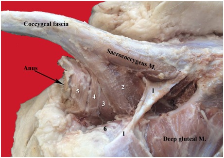 Figure 3a: A photograph the perineal region (lateral view).
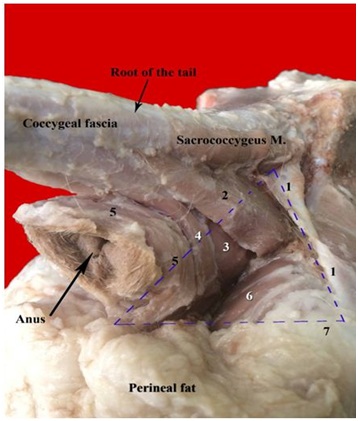 Figure 3b: A photograph showing the ischiorectal fossa (Caudolateral view).
1- Sacroischiatic ligament
2- Lateral coccygeus muscle
3- Pubococcygeus muscle
4- Ischiococcygeus muscle
5- External sphincter ani muscle
6- Internal obturator muscle
The dotted triangular region indicates the right ischiorectal fossa
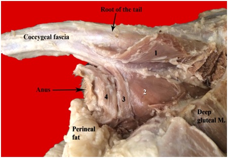 Figure 4: A photograph showing the perineal region (lateral view).
1- Lateral coccygeus muscle (reflected)
2- Pubococcygeus muscle
3- Ischiococcygeus muscle
4- External phincter ani muscle
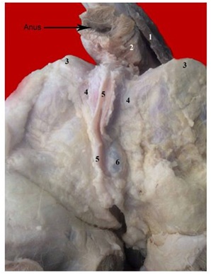
Figure 5: A photograph showing the perineal region, superficial dissection (caudal view).
1- Lateral coccygeus M
2- External phincter ani M
3- Ischial tuberosity
4- Colle’s fascia
5- Penie part of retractor penis M
6- Superficial perineal pouch
2- External phincter ani M
3- Ischial tuberosity
4- Colle’s fascia
5- Penie part of retractor penis M
6- Superficial perineal pouch
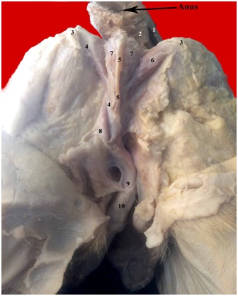 Figure 6: A photograph showing the perineal region (caudal view).
1- Lateral coccygeus M
2- External sphincter ani M
3- Ischial tuberosity
4- Colle’s fascia
5- Penile part of retractor penis M
6- Ischiocavernosus M
7- Bulbospongiosus M
8- Scrotal septum
9- Deep penile fascia
10- Superficial penile fascia
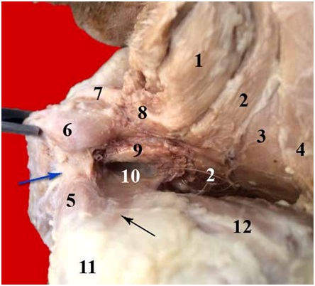
Figure 7: A photograph showing the right ischiorectal fossa (deep dissection).
1- External sphinter ani M
2- Ishiococcygeus M
3- Pubococcygeus M
4- Lateral coccygeus M
5- Ischiocavernosus M
6- Bulbospongiosus M
7- Penile part of retractor penis M
8- Perineal M
9- Pelvic urethra
10- Deep (dorsal) layer of the deep perineal fascia
11- Ischial tuberosity
12- Internal obturator M
The black arrow indicates the ischiourethralis M (covered by 10)
The blue arrow indicates superficial perineal fascia (septum between 5 and 6)
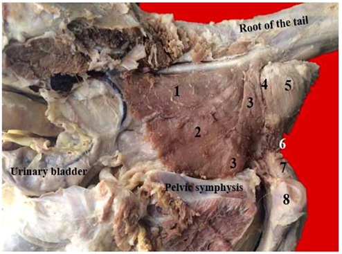 Figure 8: A photograph showing the perineal region (removed left half of pelvic bone).
1- Iliococcygeus M
2- Pubococcygeus M
3- Ischiococcygeus M
4- Vertebral part of retractor penis M
5- External sphincter ani M
6- Perineal M
7- Bulbospongiosus M
8- Left penile crus
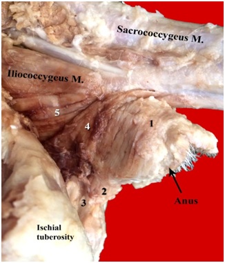 Figure 9: A photograph showing the perineal region (left lateral view).
1- External sphinter ani M
2- Perineal M
3- Perineal body
4- Ischiococcygeus M
5- Pubococcygeus M
The anal part (Suspensory apparatus of the anus), includes the rectococcygeus and the retractor penis muscle. The former, (Figures 10 and 11a) is the extrarectal continuation of the rectal musculature to the coccygeal vertebrae. The median dorsal longitudinal rectal muscle fibers coalles at the level of the pelvic out let to form a caudo dorsal projection inserted in the ventral aspect of the 5th caudal vertebra.
 Figure 10: A photograph showing the perineal region (deep dissection).
1- Retractor penis M
2- Rectum (cut)
3- Pelvic urethra
4- Prostate
5- Urinary bladder
6- Ischiococcygeus M
7- Pubococcygeus M
8- Iliococcygeus M
9- Ventral sacrococcygeus M
10- Left penile crus the yellow arrow indicates the obturator nerve
11- Rectococcygeus M
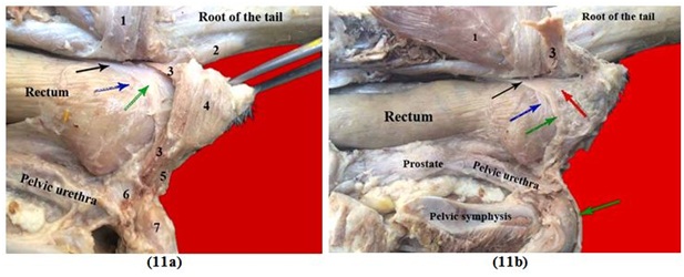
Figures 11(a,b): A photograph showing suspensory apparatus of the anus.
1- Medial coccygeus group (reflected)
2- Rectococcygeus M
3- Vertebral part of retractor penis M
4- External sphincter ani M
5- Perineal M
6- Perineal body
7- Bulbospongiosus M
The black arrow indicates the retractor penis M
The blue arrow indicates the rectal part
The green arrow indicates the penile part
The red arrow indicates the anal part of retractor penis M
The Retractor penis muscle (Figures 5,6,10,11a,11b,12), it consists of two parts; large and vertebral, the former is represented in right and left thin flat tape like in shape bundles. Each arises from the ventral aspect of the 1st and 2nd caudal vertebrae. It is directed in a caudoventral direction to run on the dorsolateral aspect of the anorectal junction. At the level of the dorsal insertion of the medial coccygeal muscle group, it inclines laterally on the anal canal. It passes on the deep face of the ischiococcygeus muscle, where the muscle divides into strong anal, penile and rectal parts. The former, is the caudal one and extends along the dorsolateral side of the anal passage, it proceeds deeply to the para-anal sinus, where the bundles terminate between the anal sphincter muscles. The penile part is the longest and crosses the lateral side of the anal canal in a vertical direction. It reaches the ventral border of the latter, where both fibers of the opposite sides are collected commonly in a single bundle and pass caudally between both perineal muscles. The muscle extends along the ventral border of penis to the glans and enrolled in the deep penile fascia. The rectal division is the cranial smaller part and its fibers are encircled with that of the terminal part of the rectum.
 Figures 12: A photograph showing the penile region, deep dissection (lateral view).
1- Deep penile fascia
2- superficial penile fascia
3- Retractor penis M
4- Ischiocavernosus M
The vertebral part (Figures 8,11a,11b), is a thin flat fascicles derive from the ventral aspect of the 3rdcaudal vertebra. The muscle passes ventrally and vertically on the caudal aspect of the external sphincter ani muscle and deeply to the ischiococcygeus muscle. The muscle fibers of both sides terminate in the bulb of penis.
The intermediate part
The perineal body is a fibrous mass ventrally located to the anal opening where the perineal fasciae attach (Figures 9 and 11a).
The perineal muscle (Figures 8,9,11a,7) is the right and left ventral median longitudinal continuation of the external sphincter ani muscle. Each short bundle ascends from the perineal body on the lateral aspects of the penile part of retractor penis muscle. The muscle bundles encircle both sides of the anal canal respectively and joined together forming the external anal sphincter.
The perineal muscle (Figures 8,9,11a,7) is the right and left ventral median longitudinal continuation of the external sphincter ani muscle. Each short bundle ascends from the perineal body on the lateral aspects of the penile part of retractor penis muscle. The muscle bundles encircle both sides of the anal canal respectively and joined together forming the external anal sphincter.
The ventral part
The perineal fasciae: The superficial perineal fascia comprises the superficial and membranous parts; the former figure 2 is a fibro-fatty layer composed of a thin fibrous sheath firmly adhered to the perineal skin and continues laterally to the superficial gluteal fascia. This layer is wrapped in fatty coat separating it from the skin externally and the membranous layer internally.
The perineal fat coat covers the ischiorectal fossae and the area of urogenital triangle, where over the former, the fat bad supports the muscles of the dorsal part of the pelvic diaphragm caudally and laterally. While on the urogenital triangle, the coat divides it from the skin externally figure 2.
The membranous layer (Colle’s fascia), (Figures 5,6,13,14) is a fibrous sheath extending from the perineal body dorsally and bilaterally stretched to the ischial tuberisities. It covers the caudoventral aspect of the ischial arch and extends ventrally to envelope the root of penis. At the arch, it coats the bulbospongiosus muscle and wraps around the penile part of retractor penis muscle. A fibrous division arises to separate between the bulbospongiosus and ischiocavernosus muscle. The fascia continues around the intrascrotal part of penis, where it receives the scrotal septum at the median plane. It proceeds cranially to join the tunica dartus of penis (superficial penile fascia) and the dorsal aspect of the latter, attached to the suspensory ligament of penis. Cranially, the dorsal extension of the superficial penile fascia, continued with the scrapa fascia of the abdominal wall. Colle’s fascia encloses a space or pouch (superficial perineal pouch) divides the deep penile fascia (internally) and perineal fat (externally). It contains the blubospongiosus and ischiocavernosus muscle.
Figures 13: A photograph showing the penile region (lateral view).
1- Suspensory ligament of penis
2- Deep penile fascia
3- Superficial penile fascia
The dotted line indicates the junction between the superficial penile fascia and scrapa
2- Deep penile fascia
3- Superficial penile fascia
The dotted line indicates the junction between the superficial penile fascia and scrapa
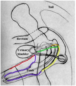 Figures 14: A diagram showing the line of extension of colle’s fascia.
The yellow lines indicates the colle’s fascia (superficial perineal fascia)
The blue line indicates the superficial penile fascia
The red line indicates the scrapa fascia of the abdominal wall
The green line indicates the perineal membrane
The brown line indicates the dorsal layer of the urogenital diaphragm
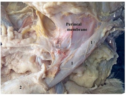
The Deep perineal fascia (Urogenital diaphragm) figures (7,12,14) is a double membranous layers that guard the ventral aspect of the pelvic outlet (Ischial arch). It is related to the prostate gland and pelvic urethra dorsally and consists of superficial (ventral) and deep (dorsal) parts. The former, is the perineal membrane that emerges from the ventral aspect of the pubic symphysis, where it sends the suspensory ligament of penis. It passes along the dorsal aspect of the root of the latter and at the ischial arch, the fascia is wrapped laterally to the ischial tuberosities. It reflects on the penile crurae and body to form the deep penile fascia. On the superficial aspect of bulbospongiosus and ischiocavernosus muscle it extends to form the colle’s fascia. The perineal membrane covers the ischiurethralis muscle laterally and proceeds cranially to the symphysis on the floor of the pelvic outlet, ventrally to the pelvic urethra to form the dorsal layer of the dorsal layer of the urogenital diaphragm. A narrow deep perineal pouch is formed between the two layers of the deep fascia and encloses the ischiocavernosus muscle figure 7.
DISCUSSION
The pelvic diaphragm was recently described within the perineal region, the present work categorized the latter into two triangular areas; anal and urogenital. The pelvic diaphragm was a group of fatty and musculofascial structures belonged to each area; a result which wasn’t recorded by the respected available literatures. The study showed the distribution of a thick fatty coat around the superficial and deep aspects of the perineum. The role of the adipose mass was to support the muscles and perineal fasciae.
In general concern, the pelvic diaphragm was integrated in three distinct regions; dorsal, intermediate and ventral. The former, represented the ischiorectal part which lodged in the anal triangle. Nearly achieved findings were mentioned by Budras et al. [12] in dog which described the ischiorectal fossa without the anal triangle. While the recent results revealed that the right and left ischiorectal fossae comprised the anal triangle. On the other hand, the intermediate perineal region connected the dorsal to the ventral one. The latter, included in the urogenital triangle and comprised the urogenital diaphragm, while the intermediate one included the perineal body and muscle. A classification which wasn’t reported in the previous available literatures. Budras et al., [12] in dog had a different classification in which they added three parts of pelvic diaphragm to the coccygeus and levator ani muscle. The authors categorized the parts into anal, perineum proper, and urogenital. The former consisted of external and internal anal muscles and rectococcygeus muscle. The proper division included the perineal body and muscle. While the urogenital one was composed of ischiocavernosus, bulbospongiosus and retractor penis muscle. In this aspect, Sisson and Grossman [13] in domestic animals, Bllenger and Canfield [9] in dog had the opinion that the pelvic diaphragm was consisted of coccygeus and levator ani muscle. The latter authors added the superficial gluteal, internal obturator, external anal sphincter muscle and sacrotuberous ligament.
It was important to note that the ischiorectal part of the pelvic diaphragm of the current study, included the coccygeus muscle group and an anal part (suspensory apparatus of anus). The former, represented the lateral coccygeus muscle and medial coccygeus group (levator ani muscle). A nearly findings were cited by Desai [14] and Miller et al. [15] in dog which reported lateral and medial coccygeus muscle and did not grouped the latter. Evans and delahunta [16] and Budras et al., [12] in dog mentioned coccygeus and levator ani muscle. While Sisson and Grossman [13] in domestic animals had the opinion that there were coccygeus and retractor ani muscle. The lateral coccygeus muscle of balady dog was described as quadrilateral in shape, arose from the ischiatic spine and had cranial, caudal, ventral and dorsal borders. Nearly recorded results were cited by Desai [14] and Miller et al., [15] in dog but they weren’t described its borders and the muscle was attached to the coccygeal vertebrae. However, the present study reported that the muscle was inserted to the lateral aspect of the first three caudal vertebrae. In this aspect, Budras et al., [11] in dog declared that the coccygeus muscle was attached to the first four caudal vertebrae. While Hall et al., [12] in dog cited that it was inserted to the first caudal vertebra.
Concerning the anatomical structures of the medial coccygeus group (levator ani muscle) in the study at hand revealed that the levator ani muscle was grouped into three muscular divisions of thin flat fascicles; Iliococcygeus, pubococcygeus and ischiococcygeus muscle. The former was the cranial and the pubococcygeus was the middle largest while the last was the caudal and smaller one. Nearly findings were cited by Koing and Liebich [17] in domestic mammals which declared that the levator ani muscle represented the iliocaudalis and ischiocaudalis muscle. On the other hand, Hall et al., [11] in dog have the opinion that there were three divisions of the levator ani muscle; pubococcygeus, iliococcygeus and puborectalis. Each part of the levator ani muscle in the balady dog had a distinct area of origin, the iliococcygeus muscle arose from the cranial border of the ilio-pubic junction and the obturator nerve separated it from the pubococcygeus muscle. The present study described the pubococcygeus muscle as the main muscle of the medial coccygeus group with fan shaped fascicles. The muscle originated from dorsal and lateral aspects of the pelvic symphysis and pubis. It was attached dorsally to the anal canal to the 4th caudal vertebra and related medially to a thick fat bad separated it from the prostate gland. A result which confermed the suggestion of Hedlund [8], Bllenger and Canfield [9], Ferreira and Delgado [6], Ribeiro [10] and Souza et al., [5] which confirmed the relation between the prostatic hyperplasia and the incidence of perineal hernia, where the hypertrophid gland will damaged the fibers of the levator ani muscle. The ischiococcygeus part of the levator ani muscle was attached from the caudo dorsal aspect of the ischial part of pelvic symphysis. In this regard, most of the available literatures, Desai [14] and Miller et al., [15] in dog, Sisson and Grossman [13] in domestic animals and Evans and delahunta [16] and Budras et al., [12] in dog had the opinion that the muscle was a single, not grouped and originated from the medial edge of ilium and dorsal aspect of the pubic symphysis. Sisson and Grossman, added that the muscle originated from the ischiatic spine and sacroischiatic ligament. The recently applied study revealed that all parts of the levator ani muscle were inserted to the 4th caudal vertebra. A little differences were recorded by Desai [14] and Miller et al., [15] in dog which determined that the muscle was inserted to the 3rd to 6th caudal vertebrae. While Budras et al., [12] in dog recorded its insertion to the 4th to 7th caudal vertebrae. The name levator ani muscle was referred to the human anatomy, as the normal attitude of the human body allowed the muscle to elevate the anus upward. Regarding that in animals, their normal body posture will changed the name to the retractor ani muscle as considered by Sisson and Grossman [13] in domestic animals. In this aspect, Drake et al., [18] in human declared that the muscle consisted of the ischiococcygeus, pubococcygeus and puborectalis. And the latter surrounded the anal opening and supported it during defecation. That was in agreement with the opinion of Hall et al., [11] in dog. While in contrary to our dissecting observation, where the parts of the medial coccygeus group were inserted to the caudal vertebrae without reinforcement fibers to the sphincter anal muscle. The ischiococcygeus muscle was separated from the external anal phincter by the vertebral head of retractor penis muscle. In this regard, the medial coccygeus muscle group of both sides formed the levator window, which allowed the anal canal to pass through freely with its suspensory apparatus. Where the study used the name medial coccygeus group rather than the levator ani one. Similarly cited results were recorded by Budras et al., [12] in dog which affirmed very few muscle fibers derived from the levator ani muscle to the external anal sphincter and the name levator ani was misleading. A results which were in contrast to that of Desai [14] and Miller et al., [15] in dog, Sisson and Grossman [13] in domestic animals which revealed that the levator ani muscle bended with the external anal sphincter. Ischiococcygeus muscle in the present dissection and that of Koing and Liebich [17] in dog was a homologous to that considered puborectalis by Drake et al., [18] in human and Hall et al., [11] in dog.
Regarding the perineal muscle in the present work, the muscle extended from the perineal body and encircled the lateral sides of the anal canal formed the external anal sphincter. Nearly results were cited by Budras et al., [12] in dog which revealed that the muscle extended from the external anal sphincter to the bulbospongiosus muscle. That was different from the opinion of Hall et al., [11] which mentioned that the anal sphincter muscle originated from the anococcygeal ligament and coursed ventrally on both sides of the external anal opening.
Our dissection revealed that the perineal body was a fibrous mass ventrally located to the anal opening where it gave an attachment for the perineal muscle and fasciae. That was different from that reported by Hall et al., [11] in dog which declared that there were no muscle fibers attached to the perineal body.
The study described the suspensory apparatus of anus and decleared that the retractor penis muscle consisted of four divisions; anal, penile, rectal and vertebral. The former was the broadest, the penile was the longest, the rectal was the smallest and all derived from a common bundle. While the vertebral division descended from the 3rd caudal vertebra. Nearly similar findings were reported by Budras et al., [12] in dog. In this regard, Evans and delahunta [16] named three parts of the muscle; pars analis, rectalis and penile.
The ventral part of the pelvic diaphragm was recently described, where it comprised the urogenital diaphragm. The latter was composed of dorsal and ventral layers which were considered the deep perineal fascia. Its dorsal layer extended from the pelvic symphysis to the level of ischial arch, where it twirled ventrally to form the ventral layer. A very small deep perineal pouch was formed between them enclosed the ischiourethralis muscle. In case of human, Drake et al., [18] recorded the external urethra muscle was wrapped in the urogenital diaphragm. The ventral layer of the urogenital diaphragm was the perineal membrane which attached cranially to the pubic symphysis. At the ischial arch, it formed the superficial perineal fascia (Colle’s fascia). A result which wasn’t cited in the available respected literatures. The Colle’s fascia covered the superficial perineal pouch which contains the bulbospongiosus and ischiocavernosus muscle. That was similar to findings that reported by Drake et al., [18] in human.
REFERENCES
- Wells DL (2007) Domestic dogs and human health: An overview. Br J Health Psychol 12: 145-156.
- Friedmann E, Son H (2009) The human- companion animal bond: How humans benefit. Vet Clin North Am Small Anim Pract 39: 293-326.
- Sjollema BE, van Sluijs FJ (1989) Perineal hernia repair in the dog by transposition of the internal obturator muscle. II. Complications and results in 100 patients. Vet Q 11: 18-23.
- Moraes PC, Facin NM, Rosa- Ballaben NM, Zanetti NM, Dias LGGG (2017) Reinforcement of the pelvic diaphragm using a purse-string suture in dogs: Description of technique. Arq Bras Med Vet Zootec 69 89-94.
- Sprada AG, Huppes RR, Feranti JPC, Souza FW, Coelho LP, et al. (2017) perineal hernia in dogs: Which technique we use? Acta Scientiae Veterinariare 45: 244.
- Ferreira F, Delgado E (2003) Hérnias perineais nos pequenos animais. Rev Port Ciênc Vet 545: 3-9.
- Costa Neto JM, Menezes VP, Toribio JMML, de Oliveira ECS, Anunciação MC, et al. (2006) Tratamento cirúrgico para correção de hérnia perineal em cão com saculação retal coexistente: (Relato de caso). Rev Bras Saúde Prod Anim 7: 7-19.
- Hedlund CS (2002) Perineal hernia. In: Fossum TW (ed.). Small animal surgery, (2nd edn). St. Louis:Mosby, Pg No: 433-437.
- Bellenger CR, Canfield RB (2003) Perineal hernia in SLATTER D. Textbook of small animal surgery (3rd edn). W B Saunders Co Ltd, Philadelphia, USA.
- Ribeiro JC (2010) Hérnia perineal em cães: Avaliação e resolução cirúrgica: Artigo de revisão. Rev Lusófona Ciênc Med Vet 3: 26-35.
- Hall MI, Plochocki JH, Sosa JR (2019) Male and female anatomical homologies in the perineum of the dog (Canis familiaris). Vet Med Sci 5: 39-47.
- Budras KD, McCarthy PH, Fricke W, Richter R (2007) Anatomy of the dog (5th edn). Schlütersche Verlagsgesellschaft mbH & Co. KG, Hannover, Germany.
- Sisson S, Grossman JD (1975) The Anatomy of the Domestic Animals (5th edn). WB Saunders Co. Philadelphia, London, Toronto.
- Desai RH (1982) An Anatomical Study of the Male and Female Pelvic Diaphragm in the Dog. University of Pennsylvania, Pennsylvania, USA.
- Evans HE (1979) Miller's Anatomy of the dog. WB Saunders Company, Philadelphia, USA.
- Evans HE, Delahunta A (2000) Guide to the dissection of the dog (5th edn). Elsevier Health Sciences, Amsterdam, Netherlands.
- König HE, Hans-Georg, Bragulla H (2014) Veterinary Anatomy of Domestic Mammals. Schattauer Verlag, Stuttgart, Germany.
- Drake R, Volg W, Mitchell A (2014) Gray’s anatomy for students (3rd edn) Elsevier, Amsterdam, Netherlands.
Citation: Nazih MA (2019) Anatomical Study on the Pelvic Diaphragm of Male Balady Dog. J Anim Res Vet Sci 3: 019.
Copyright: © 2019 Mohammed A Nazih, et al. This is an open-access article distributed under the terms of the Creative Commons Attribution License, which permits unrestricted use, distribution, and reproduction in any medium, provided the original author and source are credited.
© 2026, Copyrights Herald Scholarly Open Access. All Rights Reserved!