Antioxidant Phytochemicals Extract from L. speciosa Leaves for the Prevention and Treatment of Type 2 Diabetes Mellitus and Cardiovascular Diseases
*Corresponding Author(s):
Bidhan Chandra SarkarAssistant Professor And Chairperson, Department Of Biochemistry And Molecular Biology, Primeasia University, 12 Kemal Ataturk Avenue, Bangladesh
Tel:+88-01714063314, +880-2222275501
Email:bidhan.sarkar@primeasia.edu.bd
Abstract
Lagerstroemia speciosa. (L. speciosa) belongs to the botanical Family: Lythraceae. It is attractive and colorful pink or purple flowers. In Southeast Asia, L. speciosa is a common ornamental tree planted along roadsides, gardens, and parks. The study was carried out on the phytochemical antioxidant and antidiabetic activities of L. speciosa leaves using different solvent systems such as methanol, ethanol, acetone and n-hexane. In vitro assay, several models were used to investigate the antioxidant activity: total antioxidant capacity assay, DPPH free radical scavenging activity, ABTS free radical scavenging activity, ferrous reducing antioxidant capacity, nitric oxide scavenging activity and superoxide scavenging assay. All extracts' cytotoxic characteristics were evaluated using a brine shrimp lethality experiment. Antidiabetic activity was investigated by using Alloxan-induced diabetic rats. Out of the four extracts, the ethanol Extract of L. Speciosa Leaves (ELSL) had the highest concentrations of flavonoids (48.80 ± .37mg of Gallic acid equivalent/gm of dry extract), flavonoids (35.19 ± .71mg of Catechin equivalent/gm of dry extract) and Ethanol Extract of L. Speciosa Leaves (ELSL) possessed the highest amount of flavonols (94.44 ± .82mg of Quercetin Equivalent (QE)/gm of dry extract). Among all extracts, ELSL showed the most increased activity in ferric reducing antioxidant power assay, whereas MLSL showed maximum activity in the total antioxidant capacity assay. In DPPH, ABTS, and Nitric oxide free radical scavenging assays, ELSL showed the highest scavenging activity among all extracts with the IC50 values of 168.47μg/ml,75.78μg/ml and 141.09μg/ml, respectively. In the case of Superoxide radical scavenging assay, Methanol Extract of L. Speciosa Leaves (MLSL) exhibited maximum activity with an IC50 value of 125.42μg/ml. In brine shrimp lethality bioassay, ELSL showed the lowest cytotoxic effect with an LD50 value of 196.85μg/ml. In contrast, N-Hexane Extract of L. Speciosa Leaves (NLSL) showed the highest cytotoxic effect with an LD50 value of 119.35μg/ml.
Based on the above results (phytochemical content, antioxidant activity and cytotoxic properties), ELSL was selected to evaluate its antidiabetic activity in Alloxan monohydrate-induced diabetic rats. Compared to the diabetic control group, EB decreased the blood glucose level by 42.17% and 54.05% at doses of 100 and 200mg/kg body weight, respectively. In contrast, at the same doses, total cholesterol levels were decreased by 13.31% and 19.25%, TG reduced by 17.92% and 24.73%, LDL reduced by 40.15% and 53.09%, VLDL reduced by 17.92% and 24.83%, HDL increased by 40.43% and 45.21%. The result of this study implies that L. speciosa leaves may be good sources of antioxidants that are crucial for pharmacology and may be highly effective as a medicinal agent. Furthermore, antioxidant polyphenols could influence molecular events toward an improvement in endothelial function and, as a result, play a significant role in the prevention of cardiovascular diseases. This study is significant in preventing type 2 diabetes mellitus and cardiovascular diseases.
Keywords
Antidiabetic activity; Anti-lipidemic activity; Antioxidant activity; L. speciosa leaves extract
Introduction
Diabetes is a paired edged sword that affects both individuals and their families, and ultimately impacts on the country's economic and social development. Approximately 463 million adults (20-79 years) were living with diabetes; by 2045 this will rise to 700 million. Its caused 4.2 million deaths. Diabetes caused at least USD 760 billion dollars in health expenditure in 2019-10% of total spending on adults. In the majority of countries, an increasing number of people are being diagnosed with type 2 diabetes. 374 million peoples are at increased risk of developing type 2 diabetes (IDF Diabetes Atlas Ninth edition, 2019). Diabetes mellitus affects approximately 100 million persons worldwide[1].
This is actually the primary and effective treatment for diabetes when insulin and hypoglycemic medicines are used, but this condition also has many adverse side effects [2].Tight glycemic regulation is very well known to lower the risk of micro vascular diseases [3]. A significant number of possibly biologically active molecules, including complex carbs, alkaloids, aromatic oils, peptides, amines, hormones, phytochemicals, lipids, coumarins, sulphuric molecules and synthetic ions, have been identified [4]. There are many plants provided a full storehouse of medicines to treat all human maladies and have forms the basis of complex traditional medicine systems which have existed for thousands of years and continued to provide new solutions to mankind [5]. Common anti-diabetics plants could be a valuable source for the creation of new oral hypoglycemic substances as pharmacological entities, as simple. Looking for new antihypertensive drugs from natural plants remains desirable because they produce compounds that display effective and healthy effects on type 2 diabetes.
Phytochemicals
The potent class of substances known as phytochemicals includes a wide variety of chemical substances such polyphenols, flavonoids, steroidal saponins, organosulfur compounds, and vitamins. Phytochemicals are secondary metabolites of plants [6]. It has an important role in plant development, being part of relevant physiological process, i.e., reproduction, symbiotic association, and interactions with other organisms and the environment.
The Classification of phytochemicals
Polyphenols, terpenoids, alkaloids, phytosterols, and organosulfur compounds are the different classes of phytochemicals. The polyphenols are the most often recognized and addressed phytochemical classes [7]. There are currently 8000 polyphenolic chemicals known, and depending on their levels, they can act as both pro- and antioxidants.
Health benefit of phytochemicals
Human cells can benefit from the enormous number of phytochemicals that have recently been discovered [8, 9]. Studies shown that phytochemical-rich food has health benefits, and this supports the idea that consuming these phytochemicals might aid one's well-being [10]. Antioxidant, modern medicine throughout the world is now exposed by extensive research work on different plant species and their therapeutic concepts. The large proportion of the antioxidants separated from the larger plants were also Polyphenols. Antioxidant an effective tool has been the measurement of polyphenols and oxidative activity of dietary supplements for understanding plant medicinal property. Plants have been the source of medicinal herbal products globally for thousands of years and continue to provide humanity with new remedies approaches get an able to identify natural plant antioxidants [11]. Although the antioxidant protections vary for species to species, there is a universal existence of antioxidant defense. In the intracellular and extracellular system, antioxidants occur in both enzymatic and non-enzymatic forms [12]. Active antioxidant and free-radical scavenging activities, interest in the possible health benefits of flavonoids and other polyphenolic compounds has increased in recent years. Free radicals were molecule agents capable of rational existence, possessing one or more to mitigate damage to the cell. Moreover, some research has shown that pharmaceutical plants contain substances such as peptides, long chain fatty acids, aldehydes, flavonoids, alkaloids, essential oils, phenols, and water or organic solvent-soluble. Some compounds are significant in diagnosis for humans and animals diseases [13].
Materials and Methods
Collection of Plant Material and Authentication: For the present study Lagerstroemia speciosa were collected from the local area of Rajshahi and prove by the Department of Botany, University of Rajshahi, Rajshahi, Bangladesh. In this research works have been carried out in the Department of Biochemistry and Molecular Biology, University of Rajshahi, Bangladesh.
Preparation of plant material
The Lagerstroemia speciosa were first washed with water to remove attached dust and then chopped into tiny pieces, shed dried. The whole portion was kept grinding into a coarse powder d a grinding machine and stored in an staunch container for again use
Chemicals and apparatus
- Ethanol (Merck, Germany)
- Methanol (Sigma–Aldrich, USA)
- Acetone (Sigma-Aldrich, Germany)
- Whatman No. 1 filter paper
- N-hexene (Sigma-Aldrich,Germany)
Preparation of extract
Four different solvents namely ethanol, methanol, acetone and n-hexen were used for extraction. Around 100gm of the powdered substance was taken in different clean for each solvent round bottomed glass bottle and soaked in 500 ml of solvent. Jar was sealed with aluminium foil and silk plug kept for a period of 15 days accompanying occasional shaking and stirring.1 filter By Whatman No, the resultant extracts were filtered paper. The solvents were eventually evaporated and use a rotary evaporator in reduced pressure at 44°C. Mature, fresh leaves were collected from Lagerstroemia speciosa. After extensive washing with water the leaves were chopped into small pieces and shed dried. A high speed grinder was used for grinding the dried leaves into a coarse powder. Around 100 gm of leaves powder was soaked with 500 ml solvent and macerated for 15 days. Then the extract was collected and filtered. That filtered extract was concentrated by evaporating the solvent rotary evaporator. Obtained extract was kept in sterile glass vials and stored in refrigerator.
Phytochemical Screening of Lagerstroemia speciosa leaves extract: Phytochemical tests were carried out qualitatively for the presence of different phytochemicals like alkaloids, carbohydrates, flavonoids, glycosides, triterpenoids, resins, saponins, steroids and tannins by standard methods [14].
Reagents
- Ethanol (Merck, Germany)
- Chloroform (Merck, Germany)
- Acetone (Merck, Germany)
- Sulphuric Acid (Merck, Germany)
- Hydrochloric Acid (Merck, Germany)
- Anthrone (Sigma-Aldrich, Germany)
- Ferric Chloride
- Acetic Anhydride (Sigma-Aldrich, Germany)
- α- naphthol (Loba Chemicals, India).
Hager’stest:
A few droplets of Hager's reagent were applied to 2 mg of the extract obtained in a test tube. Yellow ppt present alkaloids.
Wagner’s test
For 1.5 percent v / v of hydrochloric acid, 2 mg of extract was acidified and a few drops of Wagner's reagent added. A brown-yellow ppt. presence of alkaloids.
Tests for carbohydrate
Anthrone test
2 mg of extract was shaken, filtered and the filtrate mixed with 10ml of water. Attached of this solution 2ml of anthrone reagent. Green or blue color formation means that carbohydrates are present.
Benedict’s test
2 mg of extract was shaken, filtered and the filtrate combined with 10ml vapor. Added to this 5 ml of the solution from Benedict and boiled for 5 minutes. Brick red colored ppt formation indicates the presence of carbohydrates
Tests for flavonoids
Shinoda’s test
2 mg of extract was dissolved in 5ml of ethanol, to which 10 drops of dilute hydrochloric acid were added accompanied by a small piece of magnesium. Presence of brown colour or pink, radish.
Tests for Glycosides
Molisch’s test
To this 2-3 drops of Molisch's reagent was applied, combined and cautiously inserted 2ml of concentrated sulfuric acid through the side of the test tube. Reddish violet ring appears, suggesting glycosides are present.
Quantitative Phytochemical Analysis
Folin-Ciocalteu method was used to estimate the total phenolic content in each extract where gallic acid is the standard.
Reagents and Apparatus
- Folin-ciocalteu reagent, FCR (Sigma-Aldrich, Germany)
- Sodium carbonate (Sigma-Aldrich, Germany)
- Methanol (Sigma-Aldrich, Germany)
- Gallic acid (Wako pure chemicals Ltd, Japan)
- Micropipette (100-1000µl)
- Pipette (1-10ml)
- UV-spectrophotometer (Shimadzu, USA)
Procedure
300 μl of extract was combined with 2.25 ml at FCR reagent at room temperature for 5 minutes, Then, added 2.25 ml Na2co3 with 90 min at room temp. Finally used at 760 nm uv spectrophotometer.The total phenolic content was expressed as equivalent in gallic acid, GAE (standard curve equation: y=.0056+.0167, R2 =.992),mg of GA/g of dry extract.
Total flavonoid test: The total amount of flavonoids was determined using the above process (Abu Bakar et al.) and the Catechin standard
Reagents and apparatus
- Aluminum chloride (Sigma-Aldrich, Germany)
- Methanol (Sigma-Aldrich, Germany)
- Catechin (Sigma–Aldrich, USA)
- UV-spectrophotometer (Shimadzu, USA)
- Sodium nitrite (Sigma-Aldrich, Germany)
- Sodium hydroxide (Sigma-Aldrich, Germany)
Procedure
Each test tube contains .5 ml of the extract mix in 0.15 ml of 5 percent sodium carbonate in followed 2.25 of d. water temperature 6 minutes. Then 0.3 ml of a 10 percent solution AlCl3.6H2O was added and allowed to stand for another 5 min before adding 1.0 ml of 1 M NaOH. Using spectro-photometer the absorbance quickly measured at 510 nm. The total flavonoid content from calibration curve was determined (standard curve equation: y= .0021x+.0352, R2=0.9916), and the results were expressed catechin equivalent per gram of dried sample (mg CA/gm).
Determination of total content in Flavonol
Maximum flavonols were estimated in the plant extracts using Kumaran and Karunakaran method.
Reagents and apparatus
- Aluminum chloride (Sigma-Aldrich, Germany)
- Sodium acetate (Sigma-Aldrich, Germany)
- Quercetin (Sigma–Aldrich, USA)
Procedure
2.0 ml of 2% AlCl3 and 3.0 ml (50g / L) sodium acetate solutions were applied to 2.0 ml sample / standard. Used 440 nm absorbance assessed at tempreture 20°C. in this ultimate concentration of 0.1 mg/ml, the standard were measured.
Determination of total proanthocyanidins
Determining the proanthocyanidin content was based on the procedure published by Sun et al.
Reagents and apparatus
- Vanillin (Sigma-Aldrich, Germany)
- Catechin (Sigma–Aldrich, USA)
Around 0.5 ml or 500 µl sample from stock were mixed with 1.5 ml vanillin solution (4%vanillin in methanol). Then 0.75ml conc. HCL was added and allowed to stand for 15 minutes at room temparature. The absorbance was quickly measured at 500 nm using spectro-photometer.
Total antioxidant capacity
With a few modifications, the Total Antioxidant Capacity (TAC) was calculated using the Prieto et al. method.
Reagents
- 6 M sulphuric acid
- 28 mM sodium phosphate: 0.397 gm sodium phosphate was dissolved in 100 ml of distilled water.
- 1% ammonium molybdate:1gm of ammonium molybdate was dissolved in 100 ml of distilled water
- Catechin (standard)
300 µl sample from stock were mixed with 3 ml reaction mixture and incubated at 95°C for 10 minutes. After cooling at room temparature, the absorbance was taken at 695nm wavelength.
Ferric reducing antioxidant power (FRAP) Test
Used by method at oyaizu (1986)
Reagents
- 1% potassium ferricyanide solution [ K3Fe(CN)6]:1gm potassium ferricyanide was dissolved into 100 ml distilled water.
- 2 M potassium buffer
- 10% Trichloroacetic Acid (TCA) solution
- 1% FeCl3 solution: 0.05gm of ferric chloride in 50 ml of distilled water was dissolved.
- Ascorbic acid
Procedure
At first each test tube added 250 µl sample from stock solution , with .625 ml of 0.2 M phosphate buffer (pH 6.6) and 1 ml of potassium ferricyanide (1%). then 20 min 50 ° C incubated adding 1 ml TCA (10 percent). Test tube, incubation mixture with 1.8 ml distilled water and 0.2 ml ferric chloride (0.1 percent). As a result, measured at 700 nm later 10 min. Improved absorption of the reaction mixture suggest greater power reduction.
Determination of DPPH free aadical scavenging activity
Reagents
- 1 mM methanol DPPH solution: 0.00394 gm DPPH has been dissolved in 100 ml of methanol.
- Methanol
- Ascorbic acid
Procedure
Various amounts of extracts were applied to a 0.1mM methanolic solution of DPPH at an equal volume (2ml). At room temperature 30 minutes absorbance recorded at 517 nm. The following formula was used to quantify radical scavenging activity:
% Scavenging Activity = (Acontrol- Asample /Acontrol) ? 100
Where, Acontrol = Absorbance of control, Asample = Absorbance of sample.
Then percentage of radical scavenging DPPH activity was plotted against concentration, and determined from graph IC50.
ABTS radical scavenging activity estimation
Based on the method described in ABTS ? + radical scavenging operation, the antioxidant capacity was determined (Re et al., 1999).
Reagents
- 7 mM 2, 2'-azino-bis (3-ethylbenzthiazoline-6-sulphonic acid) (ABTS•): 19 mg ABTS• for distilled water of 50 ml.
- Persulfate of 2.45 mMpotassium: 3.3 mg K2S2O7 per 50 ml of distilled water.
Procedure
3 ml ABTS solution was mixed with 1 ml of sample stock and stood for 6 minutes at room temparature. Then the absorbance was taken at 734 nm wavelength using spectrophotometer.
Following to formula
% Scavenging Activity = [(Acontrol– Asample) /Acontrol)]×100
Nitric oxide free radical scavenging assay
- 10mM Sodium nitroprusside
- Phosphate Buffer Saline, PBS (pH 7.4)
- Griess reagent: 1.0 ml sulfanilic acid reagent.
- Naphthylethylenediaminedihydrochloride (NED).
Procedure
2 ml of 10 mM sodium nitroprusside and 0.5 ml PBS (pH 7.4) were mixed with 0.5 ml of sample at various conc. It was incubated at 25 °C for 5 min. Then 500 µl gries reagent was added and allowed to stand for 5 minutes at room temparature followed by the addition of 1ml NED solution. That mixutre was incubated for 30 minutes at room temparature. Using spectro-photometer, the absorbance was quickly measured at 546 nm.
Superoxide free radical scavenging assay
- 50mM phosphate buffer.
- 1 mM NBT (Nitro-blue tetrazolium).
- 20µg/ml riboflavin.
- 12 mM EDTA solution.
- Ascorbic acid
Procedure
1 ml 5 Mm Na2 co3, 0.4 ml .24 Mm NBT, 2 ml 0.1 Mm EDTA, 0.4 ml 1 Mm hydroxyl amine hydrochloride were added to 500 µl sample stock and incubated for 15 minutes at 25°C. Then the absorbance was taken at 560 nm wavelength.
Brine Shrimp Lethality Bioassay
Apparatus & reagents
- Artemia salina, Leach (brine shrimp eggs)
- Sea salt (non-ionized, NaCl)
- Small tank with perforated dividing dam to hatch the shrimp
- Lamp (to atract the nauplii)
- Pipette (1 ml and 5 ml)
- Micropipette (1-100 ml adjustable)
- Glass vials (5 ml)
- Magnifying glass
Procedure
- Preparation of sea water: 38 gm NaCl (pure NaCl sigma ) dissolve in 500 ml D.H2O.
- .7gm brine eggs are added to sea water.
- O2 supply by making bubble by pump.
- Temperature maintain is very important (35-37°C).
- After 48 hrs mature naupli found /health in to water.
Preparation of sample
- 10 mg/10 ml each extract /(1mg/1ml) then this solution used as stock.
- Each test tube first fill 3ml sea water
- Then added 10 naupili count by 1000 µl micropipete with sea water.
- Added various conc. Of extracts.
- Then every test tube up to 5 ml by sea water.
- After 24 hrs count the alive naupili and use probit analysis software.
Materials
Chemicals
- Glucose estimation kit.
- Triglycerides reagent kit.
- Cholesterol kit.
- HDL cholesterol kit.
- Alloxan (Sigma–Aldrich, USA)
Working instruments
- Bio-analyzer (Micro lab, 300 -Bio-systems S.A. Barcelona, Spain)
- Eppendorf centrifuge 5415C
- Refrigerator
- Electric Balance
Plant material
Ethanol extract has been selected for the present investigation.
Test animal
Swiss albino mice were selected as experimental animal to carry out this study. Mice weighting about 20-25gm were collected from zoology department, university of Rajshahi (figure1 to 3).
 Figure 1: Normal Mice.
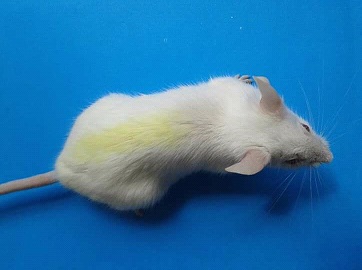 Figure 2: Diabetic mice.
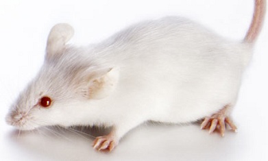 Figure 3: Treated Mice.
Induction of diabetes of mice
A single intra-peritoneal injection of alloxane (80 mg/kg body weight) into a 0.1 M sodium citrate buffer (pH-4.5) induced diabetes in overnight fasted mice. 48 hours during administration of Alloxan with a Portable Glucometer. Animals with fasting blood glucose level of approximately 11.0 mmol/L and other diabetes mellitus symptoms such as polyphagia, polydipsia, polyuria, and weight loss were deemed diabetic and included in this research.
Experimental animals grouping and treatment
The animals were divided randomly into 5 groups. Each group includes five animals (n=5). The animals have been held like this for 3 weeks:
- Group-1: Control animals feed on normal diet pellets and water.
- Group-2: An intra-peritoneal single-dose injection of 80 mg / kg body weight alloxane created the animals diabetic. Animals for whom the blood glucose levels exceeded 11.0 mmol/L at 72 h were regarded diabetic after treatment. These animals represented as undiagnosed control over diabetes.
- Group-3: The diabetic mice treated with ethanol extract of speciosa (ELSL) at a dose of 200mg/kg body weight/day for 21 days.
- Group-4: The diabetic mice treated with ELSL at a dose of 100mg/kg body/day weight for 21 days.
- Group-5: Diabetic mice were treated by Glibenclamide at a dose of 0.5mg/kg body/day weight for 21 days.
Collection and preparation of blood sample
Blood sample was collected from the tail vein for the measurement of blood glucose level on days 1, 7, 14, and 21 where participant animals were fasted overnight.
Mice were anesthetized with diethyl ether and blood was collected from the heart. Mice were sacrificed on day 21 in fasting condition. Serum Blood samples were prepared at 25°C for 15 minutes before centrifuging at 3000 rpm for 20 minutes. Finally serum was stored for further experiment carried out at -80°C in plastic vials.
Estimation of serum blood glucose
Serum glucose was measured by enzymatic colorimetric (GPO-PAP) method Micro lab, 300 (Bio-systems S.A. Barcelona, Spain).
Calculation of Result
Glucose concentration (mmol/l) = (A sample/ A standard) * 5.55
Estimation of serum Total Cholesterol (TC)
Total cholesterol (TC) was measured by enzymatic endpoint method (cholesterol Oxidase/Peroxidase) method in Micro lab, 300 (Semi autoanalyzer) using reagent of Bio Systems S.A. Barcelona Spain.
Calculation of total cholesterol result:
Cholesterol concentration (mg/dl) = (A sample/ A standard) * concentration of standard
Estimation of serum Triglycerides (TG)
Serum triglyceride was measured by enzymatic colorimetric (GPO-PAP) method Micro lab, 300 (Semiautoanalyzer, Bio Systems S.A. Barcelona, Spain).
Estimation of Serum High Density Lipoprotein (HDL)
Serum High Density Lipoprotein (HDL) was measured by enzymatic colorimetric (cholesterol CHOD-PAP) method in Micro lab, 300 (Semiautoanalyzer, Barcelona, Spain).
Calculation of LDL (serum low density lipoprotein) result
Estimation of serum low density lipoprotein (LDL) cholesterol
LDL cholesterol was determined from total cholesterol by the following formula:
Total cholesterol= HDL + LDL + (Tryglyceride / 5)
Estimation of Serum Very Low Density Lipoprotein (VLDL) Cholesterol
VLDL cholesterol was determined from total cholesterol by the following formula:
Very Low Density Lipoprotein (VLDL) = TG/5 (mg/L).
Discussion
Phytochemicals from medicinal plants have attracted immense interest as effective alternatives to pharmaceutical drugs against different diseases due to their powerful antioxidant activity. A considerable percentage of anticancer drugs used today are of plant origin. Lagerstroemia speciosa (L. speciosa), commonly known as banaba, is a deciduous tropical tree which possesses several polyphenolic compounds. The results of this investigation demonstrated that L. speciosa leaves contain substances such as alkaloid, carbohydrate, flavonoid, glycoside, resin, saponin and steroids which indicates the presence of phytochemicals in the L. speciosa leaves.
Studies suggested that the bioactivities of various plants depend on the variations in the effectiveness of the plant extract with the solvent for extraction used. Among all the tested extracts the highest amounts of total phenolic, flavonoid, flavonol and proanthocyanidins content were found in ethanol extract with the value of 48.80 ± .37 mg of Gallic acid equivalent/gm of dry extract, 35.19 ± .71 mg of Catechin equivalent/gm of dry extract, 94.44 ± .82 mg of Quercetin Equivalent (QE)/gm of dry extract and 34.8 ± 1.12 mg of Catechin/gm of dried extract respectively. Whereas, NLSL showed the lowest phenolic and flavonoid content and the lowest flavonol and proanthocyanidins content was showed by ALSL. So it can be suggested that ELSL provides more phytochemical compare to other extracts.
Many dangerous pathophysiological processes, such as cancer, diabetes and cardiovascular and neurodegenerative diseases, are associated with the accumulation of free radicals. Antioxidants neutralize the influence of free radicals, substances that harm cells in the body, and play a major role in preventing diseases. Consumption of fruits and vegetables enriched with antioxidant is known to lower the risk of several diseases caused by free radicals [15]. The use of essential oils extracted from Abrus precatorius seeds and shells have good antioxidant potential that could probably replace the use of synthetic antioxidants in further studies [16]. In this investigation, MLSL showed maximum activity in total antioxidant capacity assay and ELSL showed highest activity in ferric reducing antioxidant power assay in all concentration compare to ALSL and NLSL. The lower IC50 value indicate highest antioxidant activity. ELSL showed highest scavenging activity in DPPH, ABTS and nitric oxide radical scavenging assays among all extracts with the lowest IC50 values of 168.47μg/ml, 75.78μg/ml and 141.09μg/ml respectively. Whereas, MLSL only exhibited maximum activity in Superoxide radical scavenging assay with lowest IC50 value of 125.42 μg/ml. Thus, it can be concluded that the phenolic, flavonoid, flavonol and proanthocyanidins content present in the ELSL mostly influence the scavenging activity. Therefore, using of ELSL instead of MLSL, ALSL and NLSL is an ideal method in order to get a maximum activity of antioxidants.
The study of Waghulde et.al. suggested that alcoholic and aqueous extract of Allium fistolisum and Brassica oleraceae exhibited cytotoxic activity against the brine shrimp due to the presence of active or potent components [17]. In current investigation, LD50 is the amount of L. speciosa leaves extract given all at once that causes the 50% death of a group of brine shrimp. The value of LD50 indicates cytotoxic effect of extracts. The high lethal concentration indicates low toxicity of L. speciosa leaves extract. In this study, the ELSL content contained highest amount of lethal concentration with LD50 value of 196.85 μg/ml and NLSL showed the lowest amount of lethal concentration with LD50 value of 119.35μg/ml compare to MLSL and ALSL. So it can be concluded that ELSL is low toxic extract among others which shows low cytotoxicity.
In Southeast Asia, tea made from L. speciosa leaves has been used traditionally to treat diabetes mellitus. It has been reported recently that L. speciosa leaf extract has extensive antidiabetic, antiobesity, anti-inflammatory, antioxidant, antiviral, antibacterial, anti-hypertensive, antifibrotic and analgesic effects [18]. For the determination of antidiabetic potential of phytochemicals in vitro assay and in vivo assay were performed. In vitro assay, the study of α-amylase inhibitory activity of L. speciosa leaves extracts explained that ELSL and NLSL exhibited maximum and minimum activity respectively in any concentration among all other extracts which indicates that there is a dose dependent increase in percentage inhibitory activity against α-amylase enzyme. Thus, ELSL showed almost highest antidiabetic activity in in vitro assay.
In vivo assay, ELSL was administered to evaluate the blood glucose level of Alloxan monohydrate induced Swiss albino diabetic mice. It was found from the study that, compare to the diabetic control group, leaves extract administered diabetic mice subject showed significant reduction (P<0.001) of blood glucose level by 42.17% and 54.05% in both concentration of 100mg/kg and 200 mg/kg body weight respectively. It was also showed that the level of reduction in the blood glucose level was as near as glibenclamide administered subject which indicates the uses of ELSL instead of glibenclamide will give the same result in blood glucose reduction.
The administration of ELSL significantly reduced (P < 0.01) the serum cholesterol, LDL, VLDL and triglycerides in both concentration of 100mg/kg and 200 mg/kg body weight. Reduction of Total Cholesterol (TC) level was 13.11% (in 100 mg/kg BW) and 19.33% (in 200 mg/kg BW) observed in ELSL treated diabetic mice group whereas in positive control group reduction was 25.33%. This study also indicates that the same concentration of ELSL exhibited marked elevation in the level of HDL in the Alloxan induced diabetic group compare to the control. Therefore, the level of reduction in the serum cholesterol, LDL, VLDL and triglycerides level and the elevation of HDL was as near as glibenclamide administered subject which indicates the uses of ELSL instead of glibenclamide will give the same result in total cholesterol reduction. The aforementioned data support the assertion that L. speciosa leaves are a rich source of naturally occurring antioxidants that are clinically significant. In vitro and in vivo outcomes confirmed that the phytochemicals present on ELSL clearly shows antidiabetic effect. Moreover, these phytochemicals also act on multiple pharmacological targets to show hypolipidemic effects which is confirmed by the reduction of the total cholesterol level. Therefore, based on the result obtained from the study it is possible to say that the phenolic compounds present in the ELSL show maximum influence as therapeutic agent instead of glibenclamide which can be an ideal method in order to get recovery from type 2 diabetes mellitus and CVD with consequential health bene?ts.
Conclusion
Our study revealed that L. speciosa leaf extract has extensive antidiabetic, antioxidant and anti-hypertensive effects.
And also methanol and ethanol are preferable solvents for the extraction of polyphenols from L. speciosa leaves. Consumption of natural antioxidants may aid in maintaining an acceptable antioxidant state, minimizing oxidative stress that could cause the progression of diabetes mellitus. Among secondary metabolites, flavonoids are one of the most significant categories of bioactive chemicals. We have reported in vitro and in vivo studies the antioxidant and antidiabetic activities of some selected flavonoids. These properties of leaves extracts may contribute to the value for these plants in traditional medicine and in general medical practice. The chemical component of L. speciosa leaves extract that is responsible for the antioxidant and antidiabetic effects must thus be identified through future research.
References
- Paneni F, Beckman JA, Creager MA, Cosentino F (2003) Diabetes and Vascular Disease, Pathophysiology, Clinical Consequences and Medical Therapy: Part I. Circulation 108: 1527-1532.
- Graham TE, Wason CJ, Blüher M, Kahn BB (2007) Shortcomings in methodology complicate measurements of serum retinol binding protein (RBP4) in insulin-resistant human subjects. Diabetologia 50: 814-823.
- Gæde P, Oellgaard J, Carstensen B, Rossing P, Lund-Andersen H (2016) Years of life gained by multifactorial intervention in patients with type 2 diabetes mellitus and microalbuminuria: 21 years follow-up on the Steno-2 randomised trial. Diabetologia. 59: 2298-2307.
- Chang CL, Lin Y, Bartolome AP, Chen YC, Chiu SC (2013) Herbal therapies for type 2 diabetes mellitus: chemistry, biology, and potential application of selected plants and compounds. Evidence-Based Complementary and Alternative Medicine.
- John FN (2002) Ancient Egyptian Medicine. University of Oklahoma Press 151.
- Forni C, Facchiano F, Bartoli M (2019) Beneficial Role of Phytochemicals on Oxidative Stress and Age-Related Diseases. BioMed Research International.
- Adefegha SA, Oboh G (2011) Cooking enhances the antioxidant properties of some tropical green leafy vegetables. African Journal of Biotechnology 10: 632-639.
- Upadhyay S, Dixit M (2015) Role of Polyphenols and Other Phytochemicals on Molecular Signaling. Oxidative Medicine and Cellular Longevity .
- Budisan L, Gulei D, Zanoaga OM, Irimie AI, Sergiu C (2017) Dietary Intervention by Phytochemicals and Their Role in Modulating Coding and Non-Coding Genes in Cancer. International Journal of Molecular Sciences 18: 1178.
- Mursu J, Steffen LM, Meyer KA, Duprez D, Jacobs DR (2013) Diet quality indexes and mortality in postmenopausal women:The Iowa Women's Health Study. Am J Clin Nutr 98: 444-453.
- Krishnaiah D, Sarbatly R, Nithyanandam R (2011) A review of the antioxidant potential of medicinal plant species. Food and Bioproducts Processing 89: 217-233.
- Krishnaiah D, Bono A, Sarbatly R, Anisuzzaman SM (2013) Antioxidant activity and total phenolic content of an isolated Morinda citrifolia methanolic extract from Poly-ethersulphone (PES) membrane separator. Journal of King Saud University.
- Chandrasekhar K, Kapoor J, Anishetty S (2012) A Prospective, Randomized Double-Blind, Placebo-Controlled Study of Safety and Efficacy of a High-Concentration Full-Spectrum Extract of Ashwagandha Root in Reducing Stress and Anxiety in Adults. Indian J Psychol Med 34: 255-262.
- Shirwaikar A, Shirwaikar A, Rajendran K, Punitha ISR (2016) In Vitro Antioxidant Studies on the Benzyl Tetra Isoquinoline Alkaloid Berberine. Biol Pharm Bull 29: 1906-1910.
- Rahman MM, Islam MB, Biswas M, Alam A (2015) In vitro antioxidant and free radical scavenging activity of different parts of Tabebuia pallida growing in Bangladesh. BMC Res Notes8: 621.
- Sunday OO, Asekun OT, Familoni OB, Afolayan AJ (2014) Antioxidant and Free Radical Scavenging Capacity of Seed and Shell Essential Oils Extracted from Abrus precatorius (L). Antioxidants3: 278-287.
- Waghulde S, Kale MK, Patil VR (2019) Brine Shrimp Lethality Assay of the Aqueous and Ethanolic Extracts of the Selected Species of Medicinal Plants. Proceedings
- Mousa AM, EI-Sammad NM , AbdelHalim AH, Anwar N, Khalil WKB, et al. (2019) New-Lagerstroemia Speciosa (L.) Pers Leaf Extract Attenuates Lung Tumorigenesis via Alleviating Oxidative Stress, Inflammation and Apoptosis. Biomolecules. 9: 871.
Citation: Ahmad R, Sarkar BC, Saha HR, Dey BR, Haque S, et al. (2022) Antioxidant Phytochemicals Extract from L. speciosa Leaves for the Prevention and Treatment of Type 2 Diabetes Mellitus and Cardiovascular Diseases. J Food Sci Nutr 8: 142.
Copyright: © 2022 Rubel Ahmad, et al. This is an open-access article distributed under the terms of the Creative Commons Attribution License, which permits unrestricted use, distribution, and reproduction in any medium, provided the original author and source are credited.