Morning Glory Syndrome
*Corresponding Author(s):
Mohamed Adbellahi Cheikh AhmedDepartment Of Ophthalmology, Hospital Center Of Nouakchott, Mauritania
Tel:+222 44070724,
Email:cbadahik@gmail.com
Abstract
Morning glory syndrome is a birth defect that affects the optic nerve of the eye. The Morning Glory Syndrome (MGS) or morning glory disc anomaly was named by Kindler in 1970 because of its resemblance to the morning glory flower. Morning glory syndrome can be associated with midline cranial defects and abnormal carotid circulation. We report the case of a 10-year-old adolescent, who was brought by his parents for a profound bilateral visual acuity decrease since birth. Ophthalmic examination revealed bilateral morning glory syndrome.
Keywords
Morning glory syndrome; Morning glory disc anomaly; Congenital optic nerve dysplasia; Embryology
Introduction
Morning glory syndrome is a birth defect that affects the optic nerve of the eye. The morning glory syndrome (MGS) or morning glory disc anomaly was named by Kindler in 1970 because of its resemblance to the morning glory flower [1]. It is a congenital, funnel-shaped excavation of the posterior fundus that incorporates the optic disc, with a white tuft of glial tissue overlying the central portion of the disc and the increased number of blood vessels arising from the periphery of the disc. Morning glory syndrome can be associated with midline cranial defects and abnormal carotid circulation, such as carotid stenosis/aplasia or progressive vascular obstruction with collateralization [2].
We report the case of a 10-year-old adolescent, who was brought to a consultation by his parents for a profound bilateral visual acuity loss since birth. The patient had no notable pathological history. His Visual acuity was limited to light perception. On examination, the anterior segment and tone was normal. On the fundus, we noted in both eyes an image of morning glory around the widened papilla with vessels in the radius of a wheel and a pigmented halo within an atrophic territory. There were no associated congenital anomalies and monitoring was instituted [3] (Figures 1 & 2).
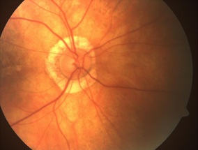 Figure 1: Right eye: morning glory disc anomaly.
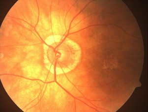 Figure 2: Left eye: morning glory disc anomaly
Conflict of interest
The authors declare no conflict of interest.
References
- Kindler P (1970) Morning glory syndrome: unusual congenital optic disk anomaly. Am J Ophthalmol 69: 376-384.
- Caprioli J, Lesser RL (1983) Basal encephalocele and morning glory syndrome. Br J Ophthalmol 67: 349-351.
- Quah BL, Hamilton J, Blaser S, Héon E, Tehrani NN (2005) Morning glory disc anomaly, midline cranial defects and abnormal carotid circulation: an association worth looking for. Pediatr Radiol 35: 525-528.
Citation: Ahmed MAC, Cheikh SS, Baba MJS (2022) Morning Glory Syndrome. J Ophthalmic Clin Res 9: 103.
Copyright: © 2022 Mohamed Adbellahi Cheikh Ahmed, et al. This is an open-access article distributed under the terms of the Creative Commons Attribution License, which permits unrestricted use, distribution, and reproduction in any medium, provided the original author and source are credited.