Patent Blue Mixed with Sodium Hyaluronate for Capsulorhexis
*Corresponding Author(s):
Fernando PolitHospital Clinica Kennedy, Guayaquil, Ecuador, United States
Tel:+593 042838636,
Email:fpolith@hotmail.com
Abstract
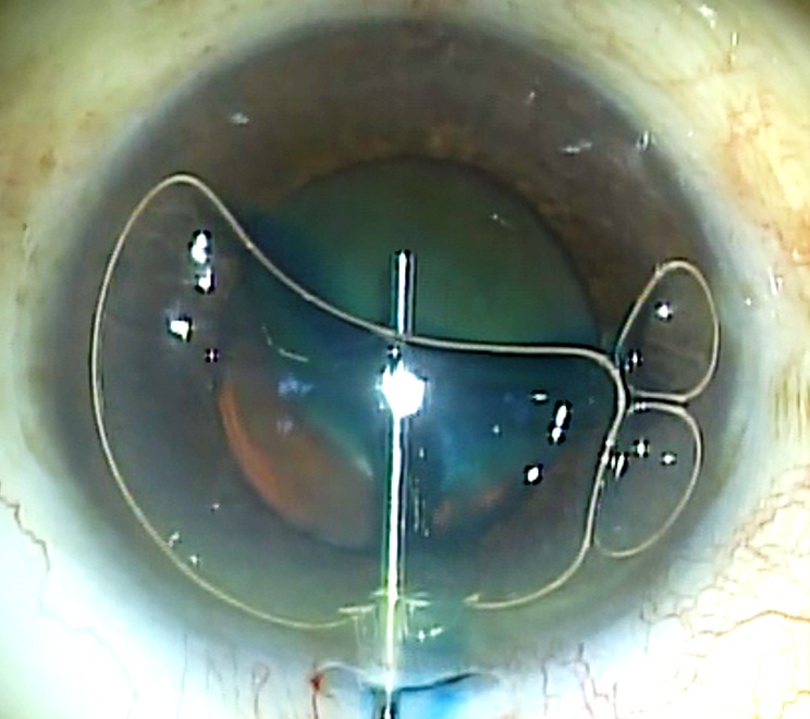
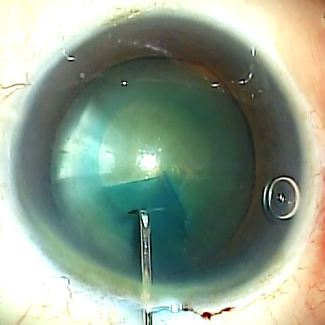
In 1999, Melles recommended the use of trypan blue dye to stain the anterior lens capsule for visualizing capsulorhexis in surgery of mature cataracts [1]. However, during the procedure the dye is in contact not only with the anterior capsule but with other anterior segment tissues, in particular the corneal endothelium. Although the complications related to the use of trypan blue have been scarce [2-5], some authors have modified the original technique to limit the dye contact to the anterior capsule [6-8].
INTRODUCTION
SURGICAL TECHNIQUE


DISCUSSION
Assisted cataract surgery femtosecond laser is currently a technique gaining users. However, the high cost of the equipment can be unaffordable for most surgeons. On the other hand, although the new technique claims higher accuracy in achieving perfect capsulorhexis, a few complications such as incomplete capsulotomy, the presence of small anterior capsular tags or even anterior radial tears have been reported [10,11]. This could explain why some surgeons still prefer to dye the anterior capsule to check the outcome of the laser assisted capsulotomy [12].
REFERENCES
- Melles GR, de Waard PW, Pameyer JH, Beckhuis HW (1999) Trypan blue capsule staining to visualize the capsulorhexis in cataract surgery. J Cataract Refract Surg 25: 7-9.
- Werner L, Apple DJ, Crema AS, Izak AM, Pandey SK (2002) Permanent blue discoloration of a hydrogel intraocular lens by intraoperative trypan blue. J Cataract Refract Surg 28: 1279-1286.
- Chowdhury PK, Raj SM, Vasavada AR (2004) Inadvertent staining of the vitreous with trypan blue. J Cataract Refract Surg 30: 274-276.
- Buzard K, Zhang JR, Thumann G, Stripecke R, Sunalp M (2010) Two cases of toxic anterior segment syndrome from generic trypan blue. J Cataract Refract Surg 36: 2195-2199.
- Burkholder B, Srikumaran D, Nanji A, Lee B, Weinberg R (2013) Inadvertent trypan blue posterior capsule staining during cataract surgery. Am J Ophthalmol 155: 625-628.
- Marques DM, Marques FF, Osher RH (2004) Three-step technique for staining the anterior lens capsule with indocyanine green or trypan blue. J Cataract Refract Surg 30: 13-16.
- Giammaria D, Giannotti M, Scopelliti A, Pellegrini G, Giannotti B (2013) Under-air staining of the anterior capsule using Trypan blue with a 30 G needle. Clin Ophthalmol 7: 233-235.
- Yetik H, Devranoglu K, Ozkan S (2002) Determining the lowest trypan blue concentration that satisfactorily stains the anterior capsule. J Cataract Refract Surg 28: 988-991.
- Kayikiçio?lu Ö, Erakgün T, Güler C (2001) Trypan blue mixed with sodium hyaluronate for capsulorhexis. J Cataract Refract Surg 27: 970.
- Bali SJ, Hodge C, Lawless M, Roberts TV, Sutton G (2012) Early experience with the femtosecond laser for cataract surgery. Ophthalmology 119: 891-899.
- Roberts TV, Lawless M, Bali SJ, Hodge C, Sutton G (2013) Surgical outcomes and safety of femtosecond laser cataract surgery: a prospective study of 1500 consecutive cases. Ophthalmology 120: 227-233.
- Hida WT, Chaves MAPD, Gonçalves MR, Tzeliks PF, Nakano CT, et al. (2014) Comparison between femtosecond laser capsulotomy and manual continuous curvilinear digital image guided capsulorrhexis. Rev Bras Oftalmol 73: 329-334.
Citation: Polit F, Polit A (2016) Patent Blue Mixed with Sodium Hyaluronate for Capsulorhexis. J Ophthalmic Clin Res 3: 021.
Copyright: © 2016 Fernando Polit, et al. This is an open-access article distributed under the terms of the Creative Commons Attribution License, which permits unrestricted use, distribution, and reproduction in any medium, provided the original author and source are credited.

