Terrien’s Marginal Degeneration with Hydrops
*Corresponding Author(s):
Anirudh DuhanSankara Eye Hospital, Pedakakani, Guntur, Andhra Pradesh, India
Tel:+91 8471873639,
Email:anirudhduhan@gmail.com
Abstract
We report a case of bilateral Terrien’s Marginal Degeneration with Hydrops. A 59 year old man presented with dimunition of vision in both eyes since 3 years. On examination both eyes had superior ectatic changes with right eye having scarred Descemet’s membrane indicating a healed hydrops and left eye showing superior corneal edema indicating resolving hydrops.
Keywords
Anterior segment; Cornea; Ectasia; Hydrops; Terrien’s
Background
Terrien’s Marginal Degeneration (TMD) is a rare, slowly progressive, peripheral corneal ectasia of unknown etiology. The condition is most commonly seen in males past the age of 40. Initially, TMD presents as small, yellow-white, stromal opacities composed of lipids with some superficial vascularization, which begins superiorly and spreads circumferentially. With progression, a gutter forms in the affected area due to stromal thinning, leaving the epithelium intact. The condition is often bilateral and asymmetric. Perforation of the cornea is rare but may occur spontaneously or secondary to trauma. Patients are often asymptomatic but may complain of mild irritation or vision changes due to increasing against-the-rule or oblique astigmatism. Very few cases have been reported of hydrops in TMD.
Case Description
A 59 year male presented with dimunition of vision in both eyes since 3 years. There was no history of trauma and no significant family history. His best corrected visual acuity was 6/24 (+5.50D/-10.0D/90) and Hand movement close to face with no improvement with glasses in right and left eye respectively. Slit lamp examination revealed superior ectatic changes in right eye with scarred Descemet’s Membrane (DM) and left eye revealed superior ectatic changes with edema. Right eye had senile immature cataract and left eye presented with senile mature cataract. Right eye fundus examination revealed no abnormalities; left eye fundus could not be viewed on slit lamp examination. B-scan ultrasound was within normal limits (Figures 1,2,3,4).
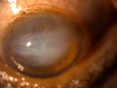 Figure 1: Right eye anterior segment OCT shows stromal scarring in the superior quadrant.
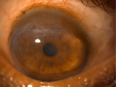 Figure 2: Left eye anterior segment OCT shows increased corneal thickness and Descemet`s detachment.
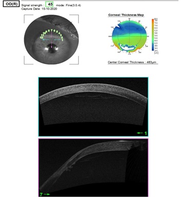 Figure 3a: Right eye showing superior superficial vascularization and scarred Descemet`s membrane depicting healed hydrops.
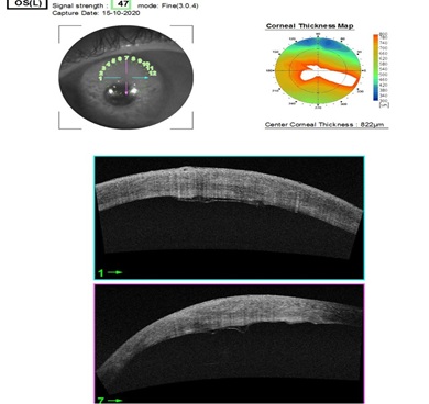 Figure 3b: Left eye showing superior superficial vascularization with stromal edema.
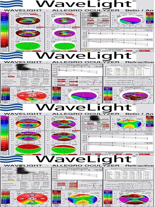
Figure 4: Pentacam topography showing steepening of anterior corneal surface with superior corneal thinning in right eye and thickening in left due to stromal edema.
Therapeutic Intervention
Patient was started on Sodium chloride (5%w/v) eye drops 5 times a day in left eye and Carboxy Methyl Cellulose (0.5%w/v) eye drops in both eyes.
Cataract surgery for left eye was advised to patient once the stromal edema reduces.
Literature Review
Terrien’s marginal degeneration is a slowly progressive thinning of the peripheral cornea.
It usually begins superior-nasally and progresses circumferentially. Yellow-white stromal opacities with superficial neovascularization are present. These stromal opacities are composed of scarring and lipid infiltration, and there is a distinct interval between the limbus and site of infiltration. Ultimately, stromal degeneration occurs, causing formation of a peripheral gutter [1]. The exact etiology of TMD is unknown, but there is male predilection (3:1). The condition usually appears in the third to fifth decade of life, but it can occur at any age [2]. Both eyes are commonly affected, but presentation can be asymmetric [1].
Discussion
Although Terrien’s marginal degeneration is rare, it is important that eycare providers differentiate it from other conditions so they can set patient expectations and initiate proper management and treatment. A thorough case history and careful slit lamp examination are essential.
Conflict of Interest
The Author declares that there is no conflict of interest.
I hereby declare that no funding was received for this work.
References
- Lopez JS, Price FW, Whitcup SM, Li Q, de Smet M, et al. (1991) Immunohistorychemistry of Terrien’s and Mooren’s corneal degeneration. Arch Ophthalmol 109: 988-992.
- Ceresara G, Migliavacca L, Orzalesi N, Rossetti L (2011) In vivo confocal microscopy in Terrien marginal corneal degeneration: a case report. Cornea 30: 820-824.
Citation: Duhan A, Sakare E, Potti S, Venigalla M (2021) Terrien’s Marginal Degeneration with Hydrops. J Ophthalmic Clin Res 8: 087.
Copyright: © 2021 Anirudh Duhan, et al. This is an open-access article distributed under the terms of the Creative Commons Attribution License, which permits unrestricted use, distribution, and reproduction in any medium, provided the original author and source are credited.