Axenfeld-Rieger Syndrome: Photo Essay
*Corresponding Author(s):
Mohamed Abdallahi Ould HamedDepartment Of Ophthalmology, Med V Military Hospital Of Rabat, Morocco
Tel:+212 655835932,
Email:badahick@gmail.com
Abstract
We report the case of a patient aged 47 who consult for a decrease in visual acuity. Examinations of the anterior segment spotting unilateral irrido-trabecular dysgenesis of the right eye with abnormal visibility of the Schwabe line corresponding to a posterior Embryotoxon and associated angular abnormalities. The eye tone measured with Goldman tonometer showed 14mmHg in the right eye and 15mmHg in the left eye. The examination of the fundus of the eye finds symmetrical morphology of the optic discs without pathological papillary excavation.
Keywords
PHOTO ESSAY
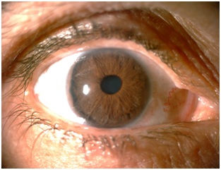
Figure 1: Embryotoxon on the temporal side of the right eye..
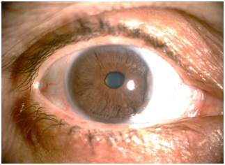
Figure 2: Embryotoxon on the nasal side of the right eye.
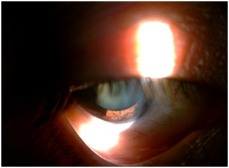
Figure 3: Angular abnormalities of the right eye.
The remainder of the somatic examination reveals no abnormality associated especially the absence of dental malformation. Chronic glaucoma is seen in 50% of patients [1].
The diagnosis of Axenfeld-Reiger syndrome uncomplicated of chronic glaucoma has been established, despite the absence of signs of Rieger [1].
No treatment has been established. Regular checks have been proposed to detect any complications including glaucoma.
This syndrome is inherited as an autosomal dominant manner. 2 genes are mainly involved in the transmission: the PITX2 gene in 4q25, present in 10-60% of patients, mainly associated with systemic alterations such as dental malformations [2,3]. The other gene responsible is FOXC1 located in 6q25, present in 50% of cases and manifested by ocular alterations, especially glaucoma [2-4].
The differential diagnosis arises with the Peters anomaly which consists of a defect of the posterior surface of the cornea associated with a stromal opacity. Currently, it is suggested that all these abnormalities are actually part of the same syndrome: Axenfeld-Rieger syndrome [5].
CONFLICT OF INTEREST
REFERENCES
- Alward WL (2000) Axenfeld-Reiger syndrome in the age of molecular genetics. Am J Ophthalmol 130: 107-115.
- Reis LM, Tyler RC, Volkmann Kloss BA, Schilter KF, Levin AV, et al. (2012) PITX2 and FOXC1 spectrum of mutations in ocular syndromes. Eur J Hum Genet 20: 1224-1233.
- Strungaru MH, Dinu I, Walter MA (2007) Genotype-phenotype correlations in Axenfeld-Rieger malformation and glaucoma patients. Invest Ophthalmol Vis Sci 48: 228-237.
- Honkanen RA, Nishimura DY, Swiderski RE, Bennett SR, Hong S, et al. (2003) A family with Axenfeld-Rieger syndrome and Peters anomaly caused by a point mutation (Phe112Ser) in the FOXC1 gene. Am J Ophthalmol 135: 368-375.
- Chang TC, Summers CG, Schimmenti LA, Grajewski AL (2012) Axenfeld-Rieger syndrome: New perspectives. Br J Ophthalmol 96: 318-322.
Citation: Hamed MAO, Soulay AY, Reda K, Oubaaz A (2018) Axenfeld-Rieger Syndrome: Photo Essay. J Ophthalmic Clin Res 5: 42.
Copyright: © 2018 Mohamed Abdallahi Ould Hamed, et al. This is an open-access article distributed under the terms of the Creative Commons Attribution License, which permits unrestricted use, distribution, and reproduction in any medium, provided the original author and source are credited.